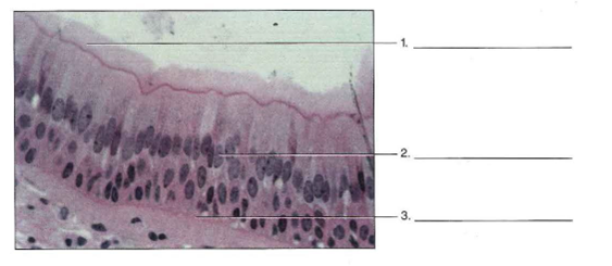
40 label the following photomicrographs by tissue type
Lab 2: Microscopy and the Study of Tissues - UW-La Crosse Epithelium is a type of tissue whose main function is to cover and protect body surfaces but can also form ducts and glands or be specialized for secretion, excretion, absorption and lubrication. Epithelial tissues are classified according to the number of cell layers that make up the tissue and the shape of the cells. Chapter 4: Tissues Flashcards | Quizlet What type of connective tissue is spring like and found in the middle walls of arteries? ... Label the following photomicrographs by tissue type and by using the terms provided. a. a. elastic fibers, b. collagenous fibers, c. reticular fibers b. a. cilia, b. nucleus, c. basement membrane
Donkey anti-Goat IgG (H+L) Cross-Adsorbed, Alexa Fluor™ 488 ... Representative photomicrographs of triple-label IF staining in LHA for Ox (Orexin-A) and GABAergic (anti-GABA) neurons.(A) 5-HT3AR, overlay image indicates an overlay of Orexin-A, 5-HT3AR and anti-GABA, (B) 5-HT1AR, overlay image indicates an overlay of Orexin-A, 5-HT1AR and anti-GABA. Confocal microscopy: Chromogens were Alexa 488 (blue ...

Label the following photomicrographs by tissue type
Tissue Identification Flashcards | Quizlet Explain how each plant tissue has a similar function to the organ or organ system in the human body. (a) dermal tissue and human skin (b) vascular tissue and the circulatory system (c) ground tissue and the skeletal system. Verified answer. BIOLOGY. Areolar Connective Tissue - Eugraph In this photo of areolar connective tissue, nuclei of cells are stained but the cytoplasm is pale and not distinguishable. As fibroblasts are the most common cells in areolar tissue, the majority of the nuclei seen here are probably fibroblast nuclei. The threadlike fibers labeled e are elastic fibers. Collagen fibers are larger in diameter ... Solved Label the following photomicrographs by tissue type | Chegg.com question: label the following photomicrographs by tissue type and by using the terms provided a. dollagenous fibers, elastic fibers, reticular fibers 2 courtesy of eric wise tissue name (400) b) basement membrane, cilia, nucleus cilia -2 nucleus - basement humbrane courtesy of eric wise tissue name (400x) c. intercalated discs, nucleus, …
Label the following photomicrographs by tissue type. Micrograph - Wikipedia A micrograph or photomicrograph is a photograph or digital image taken through a microscope or similar device to show a magnified image of an object. This is opposed to a macrograph or photomacrograph, an image which is also taken on a microscope but is only slightly magnified, usually less than 10 times. Detection of BrdU-label Retaining Cells in the Lacrimal Gland ... The photomicrographs depicted in Figure 2a-d show that following the 2- and 4-week chase periods, multiple BrdU + cells could be observed in any given lacrimal gland lobule. Figure 2a-g show that the number of BrdU + cells was high after 2 weeks of chase, decreased slightly by the 4 th week and then declined dramatically at 12 weeks. Molecular Expressions Photo Gallery: Mitosis - MagLab Mitosis is the mechanism that allows the nuclei of cells to split and provide each daughter cell with a complete set of chromosomes during cellular division. This, coupled with cytokinesis (division of the cytoplasm), occurs in all multicellular plants and animals to permit growth of the organism. In this part of the Photo Gallery, we ... LBYZOOL Act.05 Animal Histology and Organology Exercise.pdf... II. Guide Questions 1. What are the functions of the epithelium? _____ _____ _____ _____ _____ 2.
Tissues Flashcards | Quizlet Name the three types of fibers in areolar connective tissue. Collagenous, reticular, and elastic fibers What type of connective tissue is spring like and found in the middle walls of arteries? Elastic connective tissue what is the cell type in adipose tissue? loose connective tissue Name the outer connective tissue layer that envelops cartilage. identify the three phases of mitosis in the following photomicrographs Personalize learning, one student at a time. Label the figure with the following phases of the cell cycle (note the position of interphase and mitosis): • G 1 • G 2 • S • Anaphase • Metaphase • Telophase • Prophase • Cytokinesis 2. 2. In different cell types the cell cycle can last from hours to years. Tools of Cell Biology - The Cell - NCBI Bookshelf The second type of electron microscopy, scanning electron microscopy, is used to provide a three-dimensional image of cells (Figure 1.36). In scanning electron microscopy the electron beam does not pass through the specimen. Instead, the surface of the cell is coated with a heavy metal, and a beam of electrons is used to scan across the specimen. PDF Hudson City Schools / Homepage Once everyone is done, complete Qyestion #9 as a group. a. Label the apical surface on each epithelial tissue photomicrograph. b. Label the basal surface on each epithelial tissue photomicrograph. c. Draw a bracket to indicate the location of the epithelial tissue. d. Name the specific epithelial type under both tissue photomicrographs.
Chapter 6 Problem 25RQ Solution - Chegg ISBN-13: 9780077676636 ISBN: 0077676637 Authors: Eric Wise Rent | Buy. This is an alternate ISBN. View the primary ISBN for: null null Edition Textbook Solutions. RadioGraphics Describe the type of figure shown, including important imaging parameters and what can be seen on the image; Include the age, sex, and clinical history of the patient for cases of disease; For photomicrographs, include the stain and original magnification (eg, the original optical enlargement, not a recalculated value) Treatment of Figure Parts Journal of Family Medicine and Primary Care - LWW Send the images on a CD. Each figure should have a label pasted (avoid use of liquid gum for pasting) on its back indicating the number of the figure, the running title, top of the figure and the legends of the figure. Do not write the contributor/s' name/s. Do not write on the back of figures, scratch, or mark them by using paper clips. PRACTICAL 2 -TISSUES Flashcards | Quizlet In connective tissue there are more non-cellular structures present such as fibers or a matrix. Label the following photomicrographs by tissue type and by using the terms provided. a. a. elastic fibers, b. collagenous fibers, c. reticular fibers. b. a. cilia, b. nucleus, c. basement membrane.
PDF Epithelial Tissue Histology - POGIL a. Label the apical surface on each epithelial tissue photomicrograph. b. Label the basal surface on each epithelial tissue photomicrograph. c. Draw a bracket to indicate the location of the epithelial tissue. d. Name the specific epithelial type under both tissue photomicrographs. Images courtesy of The University of Kansas
identify the three phases of mitosis in the following photomicrographs The G1, S, and G2 stages are collectively known as interphase. Identify the following phases of mitosis Identify the four phases of mitosis shown in the following photomicrographs. The cell cycle consists of three major stages: 1. A. Karyokinesis (Mitosis or Nuclear Division): the cell anatomy and division.
Solved 25. Label the following photomicrographs by tissue - Chegg Label the following photomicrographs by tissue type and by using the terms provided. a. collagenous fibers, elastic fibers, reticular fibers 1. 2. 3. Courtesy of Eric Wise tissue name (400x) This problem has been solved! See the answer Show transcribed image text Expert Answer 100% (1 rating)
Blood cell histology Flashcards - Quizlet HDonica Terms in this set (4) Place the following terms and descriptions with the appropriate cell that is in the center of each of these histology slides of white blood cells. Label the types of cells in the photomicrograph using the hints provided. Label the photomicrograph using the hints provided.


Post a Comment for "40 label the following photomicrographs by tissue type"