41 art-labeling activity: structure of muscle tissues
Art-labeling Activity: Types of Connective Tissue Proper Start studying Art-labeling Activity: Types of Connective Tissue Proper. Learn vocabulary, terms, and more with flashcards, games, and other study tools. Muscles Labeling - The Biology Corner Shannan Muskopf November 11, 2020 This activity is aligned to my anatomy and physiology curriculum where students study the structure and function of muscle tissues. This has been a challenging topic to cover remotely because I can't use traditional models. Typically, I would use straws and rubber bands to model fascicles and myofibrils.
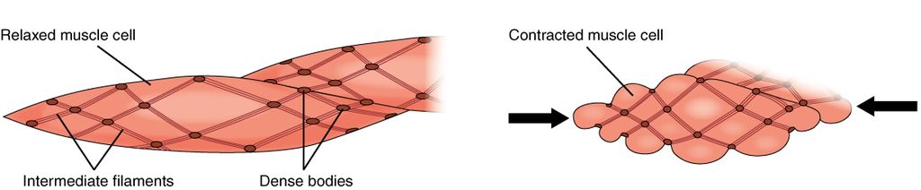
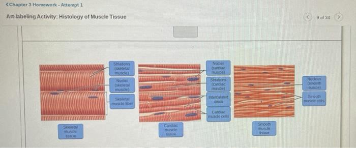
Anatomy 260 Ch3 copy.pdf - Course Hero Correct Art-labeling Activity: Histology of Nervous Tissue Label the parts of a representative neuron. Part A Drag the labels to the appropriate location in the figure. ANSWER: neural connective muscle epithelial The cell has a smooth appearance and contracts after it receives an impulse from a neuron under conscious control.
Art-labeling activity: structure of muscle tissues
Solved Secure https:/ C Lab: Histology Art-labeling | Chegg.com Answer A is skeletal mucle tissues consisting of = ENDO …. View the full answer. Transcribed image text: Secure https:/ C Lab: Histology Art-labeling Activity: Structure of muscle tissues 102091378 Part A Drag the appropriate labels to their respective targets tissue Smooth muSECM) (cardiac tissue (ECM) (skeletal Nucleus issue Type here to ... Answered: Art-labeling Activity: Structural… | bartleby Transcribed Image Text: 43 art-labeling activity: structure of muscle tissues - Product Labels ... 43 art-labeling activity: structure of muscle tissues. Oleh Angelina Zieme June 11, 2022 Post a Comment. MAP3 - Lymphatic/Immune System Flashcards | Quizlet ...
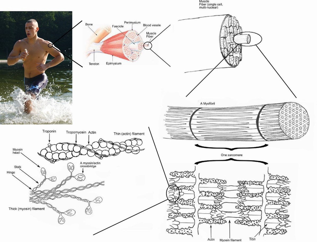
Art-labeling activity: structure of muscle tissues. Answered: Art-labeling Activity: Structural… | bartleby Q: Label different areas of an individual muscle unit known as a sarcomere below: Actin A Band Mine… A: The smallest functional unit of muscle tissue is called sarcomere. It consist of actin and myosin… Answered: Chapter 27 Homework - Altempt… | bartleby Transcribed Image Text: chapter 9- Mastering A and P, Chapter 9-1 The muscular Tissue Art-labeling Activity: The structure of a skeletal muscle fiber. PICTURE. Chapter Test - Chapter 9 Question 3 ... Art-labeling Activity: Phases and significant events of a muscle twitch in the myogram. PICTURE. ... The muscle tissue in the meat would probably not become stiff after death if it still had enough a) Calcium ... Art Labeling Activity: Layers Of The Uterine Wall - Cyclebars Beranda › Art Labeling Activity: ... the broad curved upper. Internal organs of the male reproductive system. The uterine wall is made up of three layers of muscle tissue. Ovaries and their relationship to the uterine tubes and . ... Such as samples of human speech musical tracks and illustrations of the DNA structure. Drawing Tattoo ...
Tissues Lab p5-8.pdf - Course Hero Correct The structure labeled D is a collagen fiber. Collagen fibers are thick, and appear pink due to staining. Art-Labeling Activity: Structure of muscle tissues Part A Drag the appropriate labels to their respective targets. [Solved] Art-labeling Activity: | Course Hero Answer to Art-labeling Activity: The Microscopic Structure of a Myofibril H band Zone of overlap M line A band Sarcomere Z line I band Submit Previous Answers ... Art-labeling Activity: ... A sarcomere is the area between two Z lines that can be regarded the fundamental structural and functional unit of muscle tissue. The Z-line establishes the ... sebaceous 1072017 Chapter 4 Homework Histology | Course Hero 9/14 Correct Art-Labeling Activity: Structure of muscle tissues Part A Drag the appropriate labels to their respective targets. ... ANSWER : Correct Smooth muscle tissue is found in the wall of nearly every hollow organ as well as the walls of blood vessels , the eyes , the skin , and the ducts of certain glands . ... art-labeling activity: the cell and its organelles - Blogger Smooth muscle cells are also relatively large but are long and spindle shaped. Take a toothpick and gently scrape some cells on the inside of the cheek. State a function of each organelle. Organelles are the functional parts of cellsthey are inside the cells in the cytoplasm. Sketch a model of a cell and label its parts.
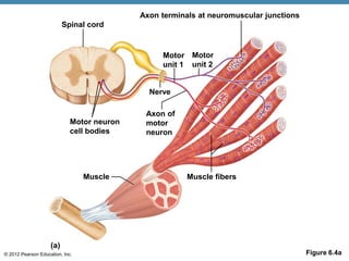
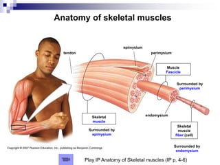
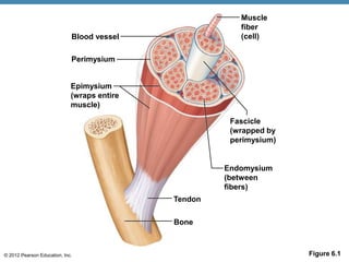
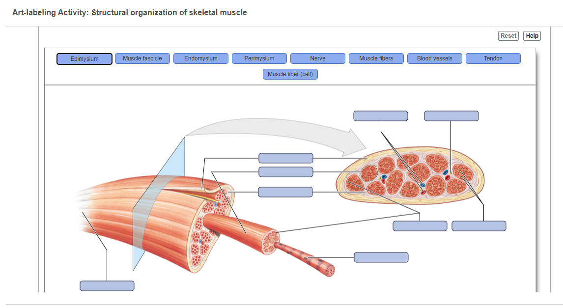
BIO 200 Chapter 9 - Muscle Tissue Physiology Flashcards - Quizlet The storage and release of calcium ions is the key function of the: sarcoplasmic reticulum. A group of skeletal muscle fibers together with the surrounding perimysium form a (n): fascicle. Art-Ranking Activity: Stages of an action potential. A crossbridge forms when: a myosin head binds to actin. A&P 1- CHAPTER 9 MASTERING ASSIGNMENTS Flashcards - Quizlet Art-labeling Activity: The structure of a skeletal muscle fiber PICTURE Which thin filament-associated protein binds two calcium ions? troponin Action potential propagation in a skeletal muscle fiber ceases when acetylcholine is removed from the synaptic cleft. Answer correct art based question chapter 4 question - Course Hero ANSWER: Correctmultinucleate cells branched cells intercalated discs situated between cells striations tendons and ligaments attached to bones heart ducts of certain glands dense irregular connective tissue smooth muscle tissue skeletal muscle tissue cardiac muscle tissue 10.2 Skeletal Muscle - Anatomy and Physiology | OpenStax These tissues include the skeletal muscle fibers, blood vessels, nerve fibers, and connective tissue. Each skeletal muscle has three layers of connective tissue (called "mysia") that enclose it and provide structure to the muscle as a whole, and also compartmentalize the muscle fibers within the muscle (Figure 10.3).
A & P Ch 6 Musclular System Student PPT
PDF Marieb HA8 chapter 4 - Pearson The word tissue derives from the Old French word meaning "to weave," reflecting the fact that the different tissues are woven together to form the "fabric" of the human body. The four basic types of tissue are epithelial tissue, connective tissue, muscle tissue, and nervous tissue. If a single, broad functional term were assigned to ...

BIO 200 Chapter 9 - Muscle Tissue Physiology Flashcards | Quizlet
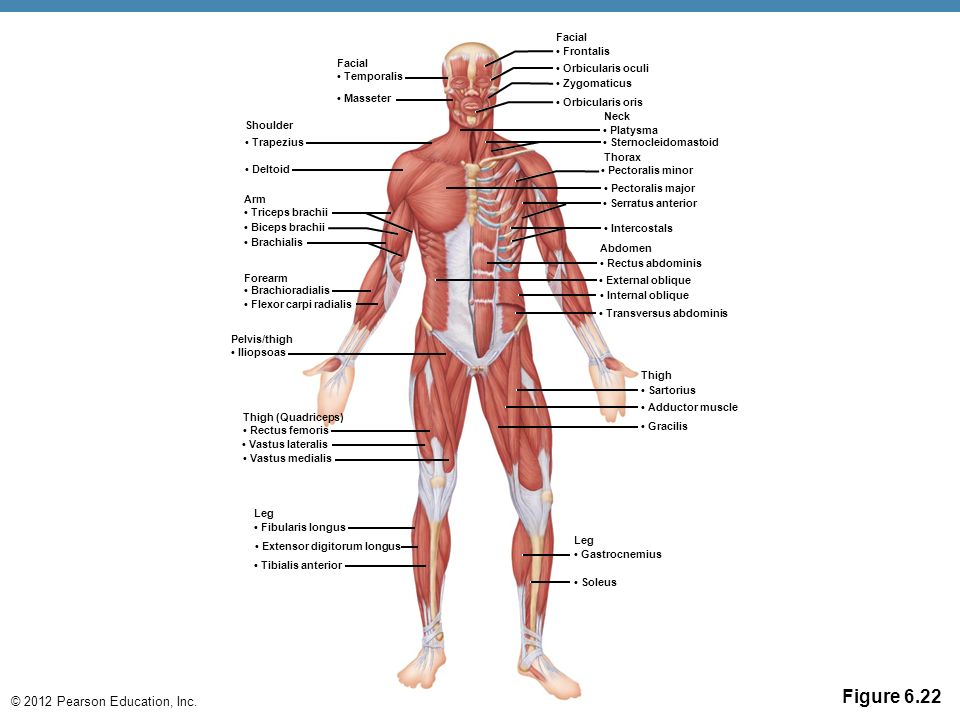
A & P Ch 6 Musclular System Student PPT - SlideShare Five Golden Rules of Skeletal Muscle Activity 1. With a few exceptions, all skeletal muscles cross at least one joint. 2. Typically, the bulk of a skeletal muscle lies proximal to the joint crossed. 3. All skeletal muscles have at least two attachments: the origin and the insertion. 4. Skeletal muscles can only pull; they never push. 5.
Skeletal muscle - Wikipedia
Answered: Art-labeling Activity: Structure of a… | bartleby Solution for Art-labeling Activity: Structure of a lymph node Medulla ... Aerobic training is any type of physical activity that uses large muscle groups and causes the body ... Match the items listed in column A with the tissue types listed in column B.Column A ... A: Cells are the basic units of life. Cells give rise to tissues and organs. ...
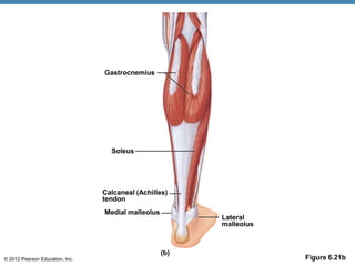
Major structures of the sole of the foot, inferior view ...
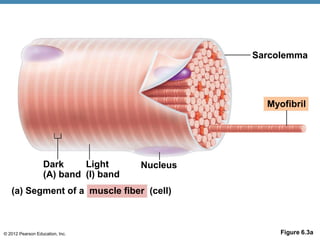
Art-labeling Activity: The Structure of a Skeletal Muscle Fiber Start studying Art-labeling Activity: The Structure of a Skeletal Muscle Fiber. Learn vocabulary, terms, and more with flashcards, games, and other study tools. ... Write. Test. PLAY. Match. Created by. BabeRuthless0504. Terms in this set (2) Art-labeling Activity: The Structure of a Skeletal Muscle Fiber... Art-labeling Activity: The Structure ...

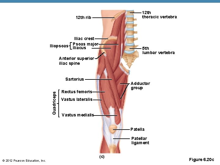
11.4 Identify the skeletal muscles and give their origins ...
Answered: Art-labeling Activity: Structure of the… | bartleby Art-labeling Activity: Structure of the testis Reset Help Seminiferous Ductus deferens tubules Epididymis Septa testis Efferent ductule Mediastinum of testis Tunica vaginalis rary Skin of scrotum Scrotal cavity Tunica On- albuginea Straight tubule Cremaster en- Lobule Dartos muscle Rete testis Expert Solution Want to see the full answer?
6 The Muscular System. - ppt video online download
Art-labeling Activity: Structure Of A Skeletal Muscle Fiber The structure of a sarcomere part a drag the labels to the appropriate location in the figure. Take the practice test transitional epithelium. Muscle tissue ( ...
12.3 Types of Muscle Tissue – Human Biology
art-labeling activity: figure 8.3 - gamezombie-danny art-labeling activity: figure 8.3 Written By tagg Tuesday, May 3, 2022 Add Comment Edit. ... Figures 8-5 and 8-6 shows many of the muscles of the bodys trunk that you need to know as well as some of the muscles of the arms and legs you will learn about in the next lab. A brand is a name picture design or symbol or combination of those items ...
Chapter 3 Homework - Attempt 1 Art-labeling | Chegg.com
43 art-labeling activity: structure of muscle tissues - Product Labels ... 43 art-labeling activity: structure of muscle tissues. Oleh Angelina Zieme June 11, 2022 Post a Comment. MAP3 - Lymphatic/Immune System Flashcards | Quizlet ...
Blog Archives - The human body
Answered: Art-labeling Activity: Structural… | bartleby Transcribed Image Text:

BIO 200 Chapter 9 - Muscle Tissue Physiology Flashcards | Quizlet
Solved Secure https:/ C Lab: Histology Art-labeling | Chegg.com Answer A is skeletal mucle tissues consisting of = ENDO …. View the full answer. Transcribed image text: Secure https:/ C Lab: Histology Art-labeling Activity: Structure of muscle tissues 102091378 Part A Drag the appropriate labels to their respective targets tissue Smooth muSECM) (cardiac tissue (ECM) (skeletal Nucleus issue Type here to ...

Skeletal Muscle | Anatomy and Physiology I

Combined pearson human anatomy ch 12 13 14 15 exam 4.pdf - 4 ...

Reproductive System Extracredit.pdf - Reproductive System ...

BIO - 168 - D6B - Nervous System Part 1 - Mastering A&P Lab ...
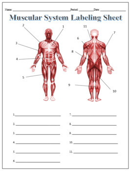
Muscular System Labeling Teaching Resources | Teachers Pay ...
A&P Muscular Chap 7.ppt

A&P 1- CHAPTER 9 MASTERING ASSIGNMENTS Flashcards | Quizlet
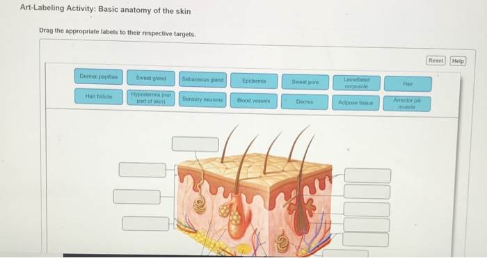
Solved Art-Labeling Activity: Basic anatomy of the skin Drag ...
Solved ▽ Part A Drag the labels onto the diagram to identify ...

Lab Exercise 12.pdf - 7/6/2021 Lab Exercise 12 Lab Exercise ...

Ch 6 Muscular System Lab quiz study practice connective ...
Bone and Cartilage

File:501 Structure of the skin.jpg - Wikimedia Commons
A & P Ch 6 Musclular System Student PPT

Chapter 21: Respiratory System (Mastering Biology) Flashcards ...
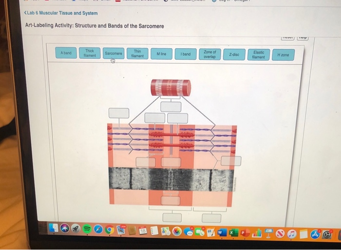
Solved (Lab 6 Muscular Tissue and System Art-Labeling | Chegg.com
A & P Ch 6 Musclular System Student PPT

Reproductive System Extracredit.pdf - Reproductive System ...
Ch 13 the peripheral nervous system and nervous activity
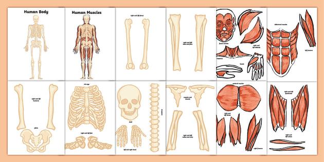
Musculoskeletal System Activity (Teacher-Made)
LAB #5

Skeletal Muscle | Anatomy and Physiology I
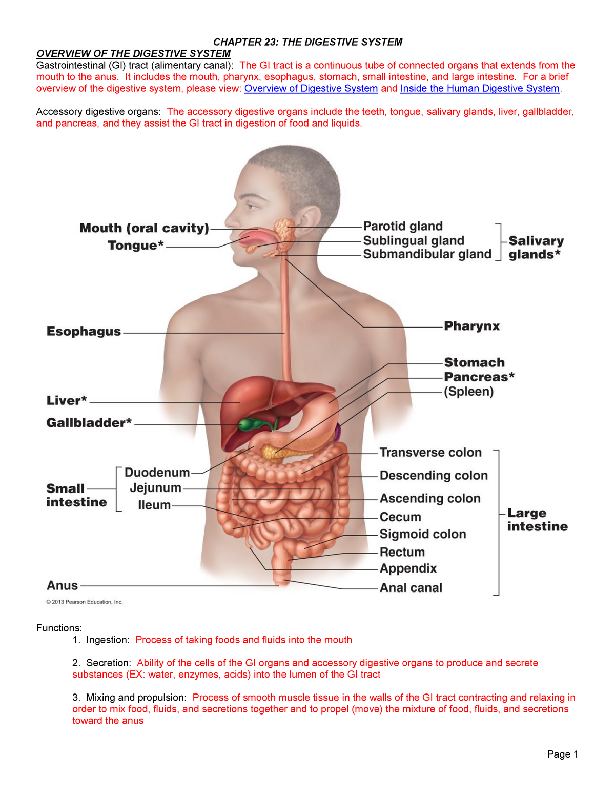
Ch. 23 Lecture Outline - OVERVIEW OF THE DIGESTIVE SYSTEM ...

Art-labeling Activity: Types of Connective Tissue Proper ...
Muscles and Muscle Tissue
Answered: Art-labeling Activity: Structural… | bartleby
![PDF] Protein synthesis rates of muscle, tendon, ligament ...](https://d3i71xaburhd42.cloudfront.net/6c60addf98287e48469b67411295eb1798cd3587/8-Figure3-1.png)
PDF] Protein synthesis rates of muscle, tendon, ligament ...
Power Point Lecture Slides Prepared by Patty BostwickTaylor
A & P Ch 6 Musclular System Student PPT

A & P Ch 6 Musclular System Student PPT
The Skeletal System The Skeletal System Components n
Post a Comment for "41 art-labeling activity: structure of muscle tissues"