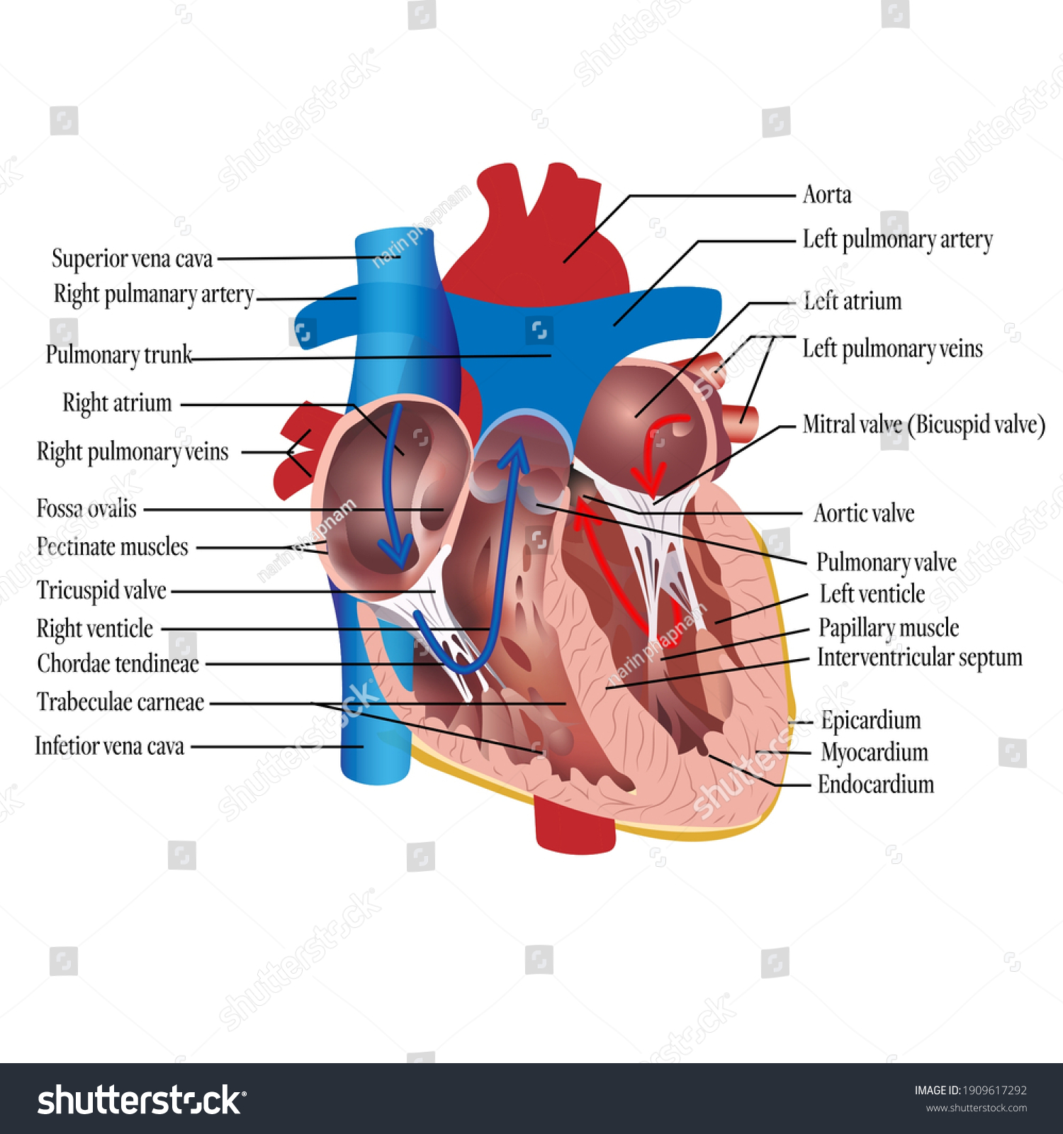
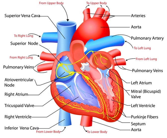
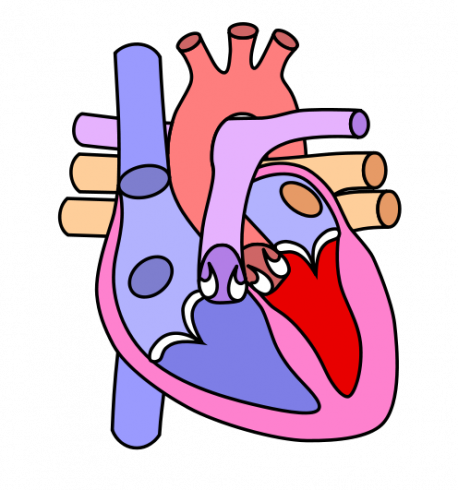
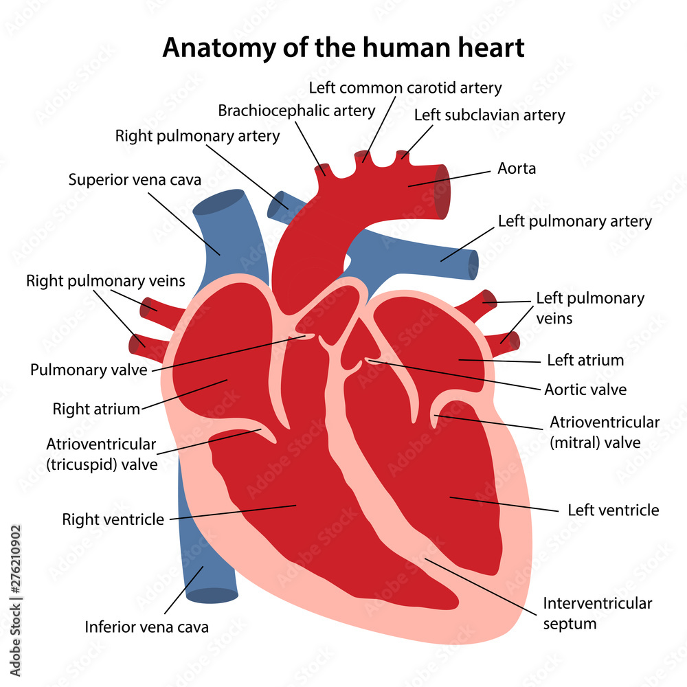
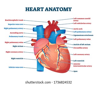
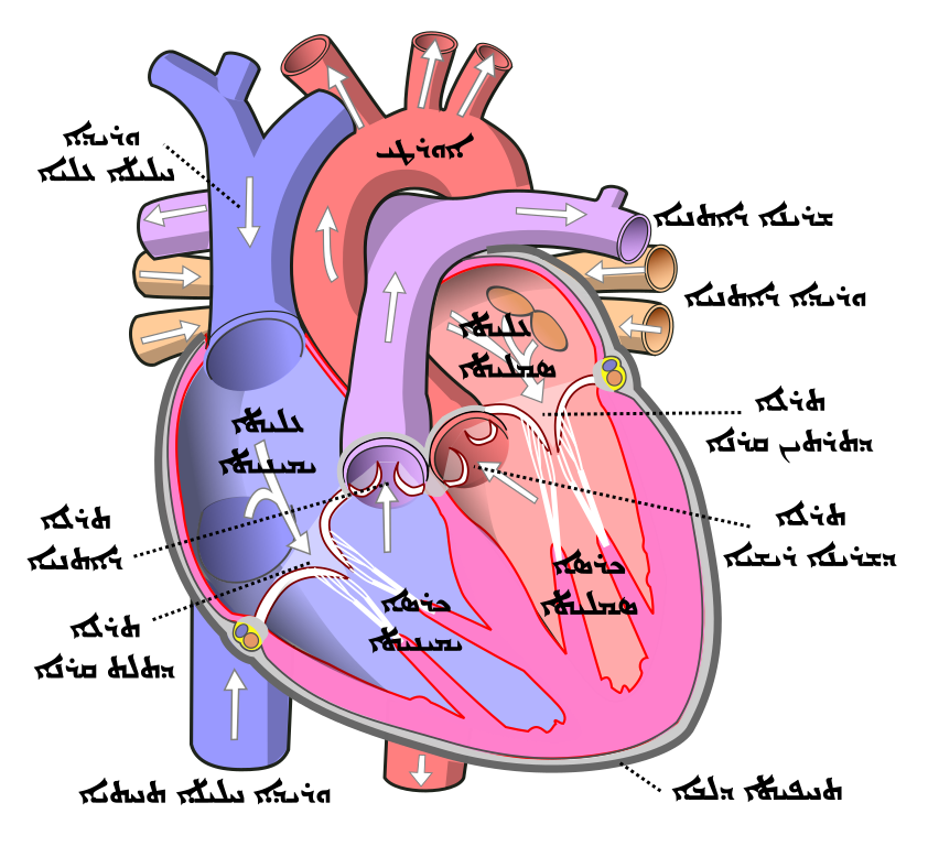
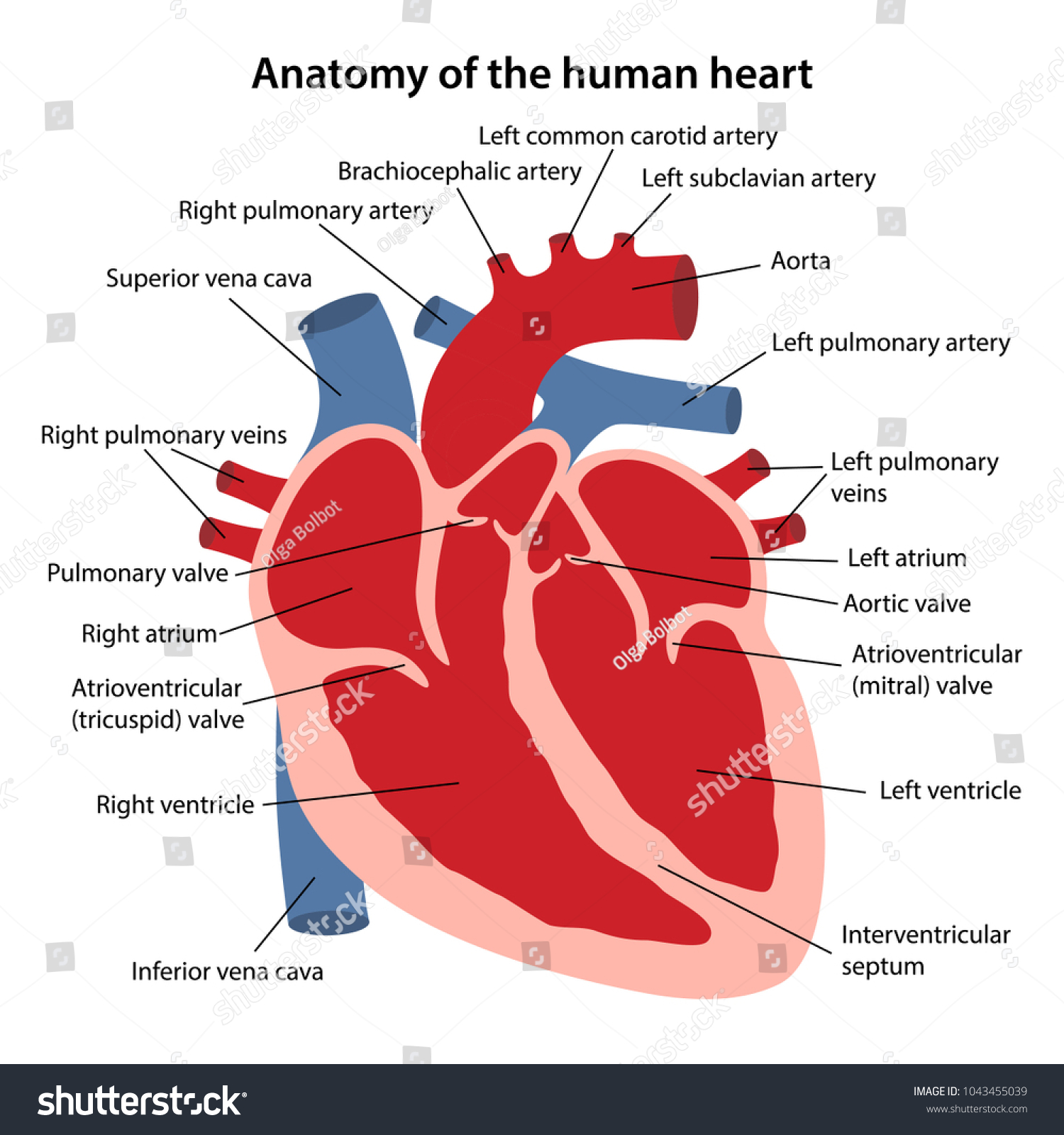
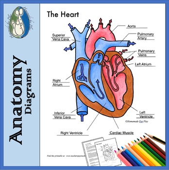
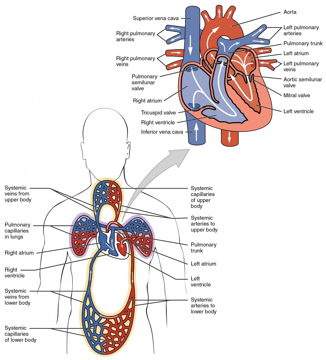
42 the human heart diagram labeled
Human eye - Wikipedia The human eye is a sensory organ, part of the sensory nervous system, that reacts to visible light and allows us to use visual information for various purposes including seeing things, keeping our balance, and maintaining circadian rhythm.. The eye can be considered as a living optical device.It is approximately spherical in shape, with its outer layers, such as the outermost, white part of ... Insulin (medication) - Wikipedia Insulin, human growth hormone (HGH) and insulin-like growth factor 1 (IGF-1) are self-administered by those looking to increase muscle mass beyond the scope offered by anabolic steroids alone. Their rationale is that since insulin and HGH act synergistically to promote growth, and since IGF-1 is a primary mediator of musculoskeletal growth, the 'stacking' of insulin, HGH …
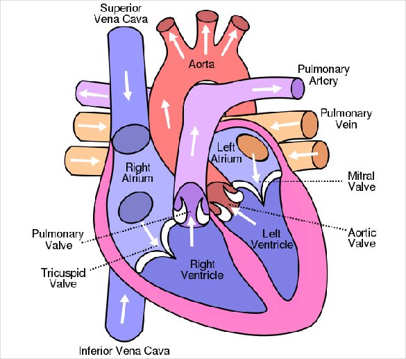
Human Heart Diagram - Side View and Top View - Heart Valve Surgery Here is a human heart diagram showing a top view of the heart - as if you were looking down on the heart. As shown below, this human heart diagram clearly illustrates the valves of the heart. The valves illustrated below are the pulmonary, tricuspid, aortic and mitral valve. So you know, I had the aortic and pulmonary valves of my heart ...

The human heart diagram labeled
Heart Diagram – 15+ Free Printable Word, Excel, EPS, PSD Teachers and students use the heart diagram, in biological science, to study the structure and functions of a human being’s heart. Friends and colleagues on the other hand may find this diagram template useful when it comes to sending special, personalized gifts to their family members and significant others. Download the template today, and ... Human Heart - Anatomy, Functions and Facts about Heart - BYJUS The human heart is divided into four chambers, namely two ventricles and two atria. The ventricles are the chambers that pump blood and atrium are the chambers that receive the blood. Among which, the right atrium and ventricle make up the "right portion of the heart", and the left atrium and ventricle make up the "left portion of the heart." 5. Muscle Anatomy Quiz - Registered Nurse RN Muscle anatomy quiz for anatomy and physiology! When you are taking anatomy and physiology you will be required to identify major muscles in the human body. This quiz requires labeling, so it will test your knowledge on how to identify these muscles (latissimus dorsi, trapezius, deltoid, biceps brachii, triceps brachii, brachioradialis, pectoralis major, serratus anterior, rectus …
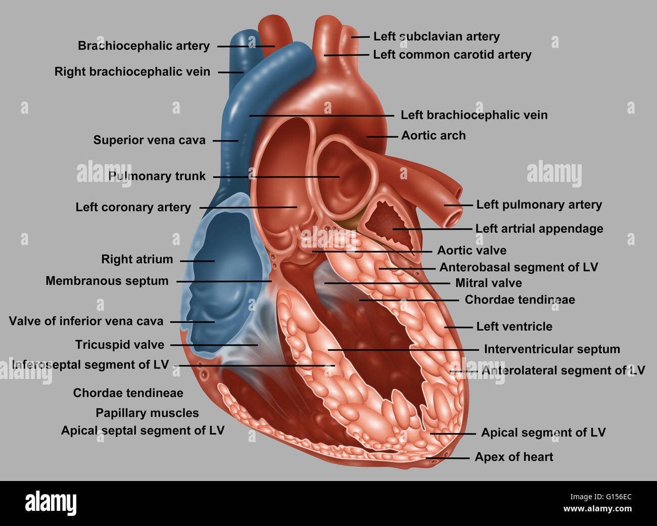
The human heart diagram labeled. heart | Structure, Function, Diagram, Anatomy, & Facts The heart consists of several layers of a tough muscular wall, the myocardium. A thin layer of tissue, the pericardium, covers the outside, and another layer, the endocardium, lines the inside. The heart cavity is divided down the middle into a right and a left heart, which in turn are subdivided into two chambers. Heart Diagram Labeled Igcse : The Human Eye Edexcel Igcse Biology ... The diagram shows a section through the human heart. The front should be labeled "blood" Sheep heart dissection lab anatomy & It is an interactive, lecture based course covering the underlying concepts and principles related to human gross anatomy of the heart and related structures. Heart Diagram with Labels and Detailed Explanation - BYJUS Diagram of Heart. The human heart is the most crucial organ of the human body. It pumps blood from the heart to different parts of the body and back to the heart. The most common heart attack symptoms or warning signs are chest pain, breathlessness, nausea, sweating etc. The diagram of heart is beneficial for Class 10 and 12 and is frequently ... The Anatomy of the Heart, Its Structures, and Functions - ThoughtCo The heart is the organ that helps supply blood and oxygen to all parts of the body. It is divided by a partition (or septum) into two halves. The halves are, in turn, divided into four chambers. The heart is situated within the chest cavity and surrounded by a fluid-filled sac called the pericardium. This amazing muscle produces electrical ...
Anatomy of a Human Heart - U of M Health Located between the lungs in the middle of the chest, the heart pumps blood through the network of arteries and veins known as the cardiovascular system. It pushes blood to the body's organs, tissues and cells. Blood delivers oxygen and nutrients to every cell and removes the carbon dioxide and other waste products made by those cells. Diagram of Human Heart and Blood Circulation in It Exterior of the Human Heart A heart diagram labeled will provide plenty of information about the structure of your heart, including the wall of your heart. The wall of the heart has three different layers, such as the Myocardium, the Epicardium, and the Endocardium. Here's more about these three layers. Epicardium Label the heart — Science Learning Hub Label the heart Interactive Add to collection In this interactive, you can label parts of the human heart. Drag and drop the text labels onto the boxes next to the diagram. Selecting or hovering over a box will highlight each area in the diagram. pulmonary vein semilunar valve right ventricle right atrium vena cava left atrium pulmonary artery Human Heart Anatomy (3D Medical Animation) - YouTube Using state of the art 3D animation techniques, this video shows the anatomy of the heart. Includes close ups of the superior vena cava, rights and left atrium, the valvles, the ventricles and the...
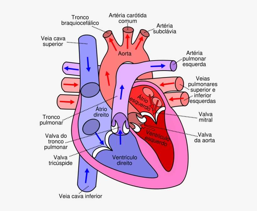
Diagrams, quizzes and worksheets of the heart | Kenhub Using our unlabeled heart diagrams, you can challenge yourself to identify the individual parts of the heart as indicated by the arrows and fill-in-the-blank spaces. This exercise will help you to identify your weak spots, so you'll know which heart structures you need to spend more time studying with our heart quizzes. A Labeled Diagram of the Human Heart You Really Need to See The human heart, comprises four chambers: right atrium, left atrium, right ventricle and left ventricle. The two upper chambers are called the left and the right atria, and the two lower chambers are known as the left and the right ventricles. The two atria and ventricles are separated from each other by a muscle wall called 'septum'. Human Heart - Diagram and Anatomy of the Heart - Innerbody The inferior tip of the heart, known as the apex, rests just superior to the diaphragm. The base of the heart is located along the body's midline with the apex pointing toward the left side. Because the heart points to the left, about 2/3 of the heart's mass is found on the left side of the body and the other 1/3 is on the right. A Diagram of the Heart and Its Functioning Explained in Detail The heart blood flow diagram (flowchart) given below will help you to understand the pathway of blood through the heart.Initial five points denotes impure or deoxygenated blood and the last five points denotes pure or oxygenated blood. 1.Different Parts of the Body ↓ 2.Major Veins ↓ 3.Right Atrium ↓ 4.Right Ventricle ↓ 5.Pulmonary Artery ↓ 6.Lungs
Heart Blood Flow | Simple Anatomy Diagram, Cardiac Circulation ... - EZmed Diagram: Blood flow through the heart in 12 steps, cardiac anatomy and structures, and cardiac circulation pathway. Blood Flow - Right Side of the Heart Let's now walk through the above 12 steps beginning with the right side of the heart. 1. Superior Vena Cava and Inferior Vena Cava
Heart Pictures, Diagram & Anatomy | Body Maps - Healthline The average human heart weighs between 6 and 11 ounces. The muscle is strong enough to pump up to 2,000 gallons — as much as a fire department's tanker truck — of blood through one's body ...
Human Heart Diagram Class 10 | Get Easy Tricks to Draw Human Heart Human Heart Diagram Class 10. When in class 10th, we are required to draw the human heart in exams as well. This can get even more stressful if it is the final/ board exam. It is always an important essay-type question that carries high weightage in terms of marks. So, you need to practice these multiple times.
Male Human Anatomy Diagram Pictures, Images and Stock Photos Labeled human anatomy diagram of man's full body muscular system from a posterior view on a white background. Cardiovascular System The human cardiovascular system (also known as the circulatory system). A medical diagram showing the heart, arteries and veins of the human body. male human anatomy diagram stock illustrations . Cardiovascular System. The human …
How the Heart Works: Diagram, Anatomy, Blood Flow - MedicineNet The heart is located under the rib cage -- 2/3 of it is to the left of your breastbone (sternum) -- and between your lungs and above the diaphragm. The heart is about the size of a closed fist, weighs about 10.5 ounces, and is somewhat cone-shaped. It is covered by a sack termed the pericardium or pericardial sack.
Human Heart (Anatomy): Diagram, Function, Chambers, Location in Body The heart is a muscular organ about the size of a fist, located just behind and slightly left of the breastbone. The heart pumps blood through the network of arteries and veins called the...
Male anatomy drawing labeled 2022. 7. 1. · Anatomical Drawings Of A Fetal Pig blog.valdosta.edu. pig fetal drawing heart anatomy vessels labeled internal diagram blood dissection drawings identify following use male anatomical important paintingvalley. Duke Anatomy - Lab 2 Optional Exercise web.duke.edu. anatomy surface duke optional exercise lab atlas. Dissection Of The Fetal Pig. Line Drawing Of …
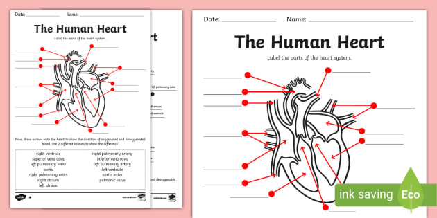
The Human Heart Labeling Worksheet (Teacher-Made) - Twinkl The human heart is a muscle made up of four chambers, these are: Two lower chambers - the left and right ventricles. It's also made up of four valves - these are known as the tricuspid, pulmonary, mitral and aortic valves. With this heart diagram without labels, you can familiarise your students with all the correct terms and help them ...
Human Heart Diagram Labeled | Science Trends Human Heart Diagram Labeled Daniel Nelson 1, January 2019 | Last Updated: 3, March 2020 The human heart is an organ responsible for pumping blood through the body, moving the blood (which carries valuable oxygen) to all the tissues in the body. Without the heart, the tissues couldn't get the oxygen they need and would die.
Anatomy of the human heart - Wikipedia The heart has the shape of a pyramid, with its apex pointing towards the left nipple while its base forms the posterior surface of the heart. Other surfaces are the anterior, inferior (or diaphragmatic), and two pulmonary surfaces facing the lungs.
14,796 Human heart diagram Images, Stock Photos & Vectors - Shutterstock Human heart diagram royalty-free images 14,796 human heart diagram stock photos, vectors, and illustrations are available royalty-free. See human heart diagram stock video clips Image type Orientation Color People Artists Sort by Popular Anatomy Healthcare and Medical Abstract Designs and Shapes heart medicine organ circulatory system hemodynamics
Heart Anatomy: Labeled Diagram, Structures, Blood Flow ... - EZmed There are 4 chambers, labeled 1-4 on the diagram below. To help simplify things, we can convert the heart into a square. We will then divide that square into 4 different boxes which will represent the 4 chambers of the heart. The boxes are numbered to correlate with the labeled chambers on the cartoon diagram. View fullsize
Learn all muscles with quizzes and labeled diagrams | Kenhub 14.09.2022 · How to learn all muscles with quizzes and labeled diagrams Author: Molly Smith DipCNM, mBANT • Reviewer: Dimitrios Mytilinaios MD, PhD Last reviewed: September 14, 2022 Reading time: 3 minutes So, you want to learn about human muscle anatomy. But where to start?! The number of muscles of the body is estimated to be around 600, so there’s ...
Labeled Skeletal System Diagram - Bodytomy A basic human skeleton is studied in schools with a simple diagram. It is also studied in art schools, while in-depth study of the skeleton is done in the medical field. This article explains the bone structure of the human body, using a labeled skeletal system diagram and a simple technique to memorize the names of all the bones.
Label the Heart Diagram | Quizlet Start studying Label the Heart. Learn vocabulary, terms, and more with flashcards, games, and other study tools.

Diagram of heart/How to draw human heart easily/human heart/human heart diagram/labelled human heart
Diagram of the human heart royalty-free images - Shutterstock 14,830 diagram of the human heart stock photos, vectors, and illustrations are available royalty-free. See diagram of the human heart stock video clips. Image type.
Organs Of The Human Body Diagram stock illustrations Detailed human body diagram. Each element grouped and labeled for easy use and editing. sympathetic and parasympathetic nervous system 3d medical vector illustration on white background sympathetic and parasympathetic nervous system 3d medical vector illustration on white background eps 10 organs of the human body diagram stock illustrations . sympathetic …
label heart diagram worksheet Related Items. 9 Pictures about Related Items : Heart Diagram Worksheet Blank Human Heart Worksheets Label Heart, Label the heart worksheets (SB6634) - SparkleBox and also Well Labelled Diagram Of Human Digestive System - Made By Creative Label.
Diagram of Blood Flow Through the Heart - Bodytomy If not, you can have a look at the labeled diagram of the human heart present in this article. The strongest muscle in the human body is the human heart. The human heart continues to pumps liters of blood throughout the body all lifelong. The minute the heart stops pumping blood, it leads to a heart attack that can prove to be fatal. Blood Circulation within the Heart for Kids. The …
Heart: Anatomy and Function - Cleveland Clinic Heart. Your heart is the main organ of your cardiovascular system, a network of blood vessels that pumps blood throughout your body. It also works with other body systems to control your heart rate and blood pressure. Your family history, personal health history and lifestyle all affect how well your heart works. Appointments 800.659.7822.
Muscle Anatomy Quiz - Registered Nurse RN Muscle anatomy quiz for anatomy and physiology! When you are taking anatomy and physiology you will be required to identify major muscles in the human body. This quiz requires labeling, so it will test your knowledge on how to identify these muscles (latissimus dorsi, trapezius, deltoid, biceps brachii, triceps brachii, brachioradialis, pectoralis major, serratus anterior, rectus …
Human Heart - Anatomy, Functions and Facts about Heart - BYJUS The human heart is divided into four chambers, namely two ventricles and two atria. The ventricles are the chambers that pump blood and atrium are the chambers that receive the blood. Among which, the right atrium and ventricle make up the "right portion of the heart", and the left atrium and ventricle make up the "left portion of the heart." 5.
Heart Diagram – 15+ Free Printable Word, Excel, EPS, PSD Teachers and students use the heart diagram, in biological science, to study the structure and functions of a human being’s heart. Friends and colleagues on the other hand may find this diagram template useful when it comes to sending special, personalized gifts to their family members and significant others. Download the template today, and ...
















Post a Comment for "42 the human heart diagram labeled"