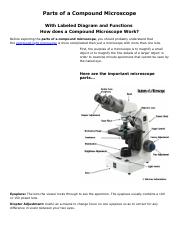
45 compound microscope diagram labeled
Compound Microscope: Definition, Diagram, Parts, Uses, Working ... - BYJUS The parts of a compound microscope can be classified into two: Non-optical parts Optical parts Non-optical parts Base The base is also known as the foot which is either U or horseshoe-shaped. It is a metallic structure that supports the entire microscope. Pillar The connection between the base and the arm are possible through the pillar. Arm Parts of a Microscope and Their Functions - Microbiology Note Jul 03, 2020 · microscope labeled diagram | Source: Microbiologynote.com. The microscope parts are divided into two main categories, such as; Structural parts of microscope ; ... Compound Microscope: This word was once used to describe a microscope with more than one objective lens. Now, most people think of a compound microscope as a high-power microscope ...
Diagram of a Compound Microscope - Biology Discussion The size of objects viewed under the compound microscope can be accurately determined using a micrometer. The latter consists of two scales, the eyepiece scale, (also called 'graticule' or 'ocular') and the stage micrometer scale. The eyepiece scale is calibrated with the help of stage micrometer and the former is then used for measurements.

Compound microscope diagram labeled
Labelled Diagram of Compound Microscope The below mentioned article provides a labelled diagram of compound microscope. Part # 1. The Stand: The stand is made up of a heavy foot which carries a curved inclinable limb or arm bearing the body tube. The foot is generally horse shoe-shaped structure (Fig. 2) which rests on table top or any other surface on which the microscope in kept. Label a Compound Microscope Diagram | Quizlet Start studying Label a Compound Microscope. Learn vocabulary, terms, and more with flashcards, games, and other study tools. ... Label this. Illuminator Switch. Sets found in the same folder. A&P II Ch. 24 Digestive Lab QUIZ. 10 terms. CWRN2016. Body planes Label. 9 terms. Hesi_Study. Labeled Parts Compound Microscope [4THWBQ] A compound light microscope is a type of light microscope that uses a compound lens system meaning it operates through two sets of lenses to magnify the image of a specimen Two different compound light microscope models with their parts labeled Leica DM1000 Fluorescence Filter - Blue - 11513828 Compound Microscopes Defining Features Image 1 ...
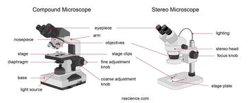
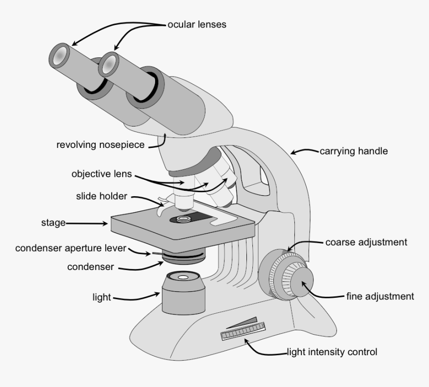
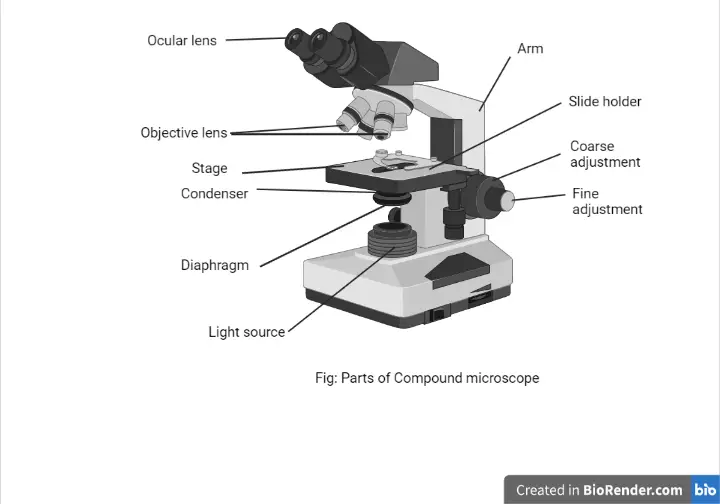
Compound microscope diagram labeled. Compound Microscope- Definition, Labeled Diagram, Principle, … Apr 03, 2022 · Parts of a Compound Microscope. Eyepiece And Body Tube. The eyepiece is the lens through which the viewer looks to see the specimen. It usually contains a 10X or 15X power lens. The body tube connects the eyepiece to the objective lenses. Objectives and Stage Clips. Objective Lenses are one of the most important parts of a Compound Microscope. Multiple Alleles: Definition, Types, and Examples - Research Tweet Aug 27, 2021 · This hybrid, also known as the vestigial antlered compound, is made up of two mutant genes at the same locus. There is evidence of Mendelian segregation and recombination. ... Microscope, Microscope Parts, Labeled Diagram, and Functions September 3, 2022 View Details » Pinocytosis: Definition, Types, Features, and Functions June 29, 2022 A Study of the Microscope and its Functions With a Labeled Diagram ... To better understand the structure and function of a microscope, we need to take a look at the labeled microscope diagrams of the compound and electron microscope. These diagrams clearly explain the functioning of the microscopes along with their respective parts. Man's curiosity has led to great inventions. The microscope is one of them. Parts of Stereo Microscope (Dissecting microscope) – labeled diagram ... If you would like to learn optical components of a compound microscope, please visit Compound Microscope Parts – Labeled Diagram and their Functions, and this article. How to use a stereo (dissecting) microscope. Follow these steps to put your stereo microscopes in work: 1. Set your microscope on a tabletop or other flat sturdy surface where ...
What is a Stereo Microscope? - New York Microscope Company May 11, 2018 · Each part of a stereo microscope is labeled in the diagram below. This example is a typical classroom type stereo microscope with track stand and built-in illumination. ... A compound microscope helps to look at samples under magnifications as high as 40x to 1000x or more. Examples include the viewing of animal or plant cells, blood counts ... Compound Microscope Parts – Labeled Diagram and their ... Labeled diagram of a compound microscope Major structural parts of a compound microscope There are three major structural parts of a compound microscope. The head includes the upper part of the microscope, which houses the most critical optical components, and the eyepiece tube of the microscope. Light Microscope- Definition, Principle, Types, Parts ... Sep 07, 2022 · Brightfield Light Microscope (Compound light microscope) This is the most basic optical Microscope used in microbiology laboratories which produces a dark image against a bright background. Made up of two lenses, it is widely used to view plant and animal cell organelles including some parasites such as Paramecium after staining with basic stains. microscope worksheet label microscope parts compound worksheet diagram printable worksheets quiz grade biology labeling science answers sketch labeled tests blank light microscopio label. ... Microscope parts labeled diagram worksheet label lens worksheeto via. Label the microscope by crista tiboldo. Microscope compound parts light worksheet diagram labeled worksheeto via
Microscope, Microscope Parts, Labeled Diagram, and Functions Sep 03, 2022 · Simply multiply the magnification of the ocular lens by the magnification of the objective lens to calculate the power of magnification of a microscope. For a typical compound microscope with a 10X ocular lens and objective lenses with magnifications of 4X, 10X, 40X, and 100X, your microscope will have magnifications of 40X, 100X, 400X, and ... 16 Parts of a Compound Microscope: Diagrams and Video In compound microscopes with two eye pieces there are prisms contained in the body that will also split the beam of light to enable you to view the image through both eye pieces. 2. Arm The arm of the microscope is another structural piece. The arm connects the base of the microscope to the head/body of the microscope. Plant Tissue Culture Laboratory (With Diagram) - Biology … Microscope: Simple, compound, inverted binocular dissection microscopes are essential for various purposes. Some of the microscopes (Figs 1.3 and 1.4) should be fitted with a camera for taking photomicrograph. Microtome: It is needed for sectioning the cultured tissue. Wooden Rocks: These are required for keeping the various chemicals. Compound Microscope Labeled Diagram | Quizlet Compound Microscope Labeled Diagram | Quizlet Compound Microscope Labeled + − Flashcards Learn Test Match Created by meganplocher734 Terms in this set (14) Eyepiece/Ocular lens Contains the ocular lens Body tube A hollow cylinder that holds the eyepiece. Arm Part that supports the microscope. Stage Supports the slide or specimen
2.1 " Compound Microscope" | Download Scientific Diagram
Cell membrane - definition, structure, function, and biology - Rs' … [In this figure] The anatomy of an animal cell with organelles labeled. ... Schematic diagram of transmembrane, peripheral, and lipid-anchored membrane proteins. ... Under a compound light microscope, the cell membrane (only 5-10 nm) may be too thin to be seen. However, you can easily tell the boundary of cells if stained with proper dyes.

List: Parts of a Microscope and their Function | Pathwooded
Compound Microscope - Diagram (Parts labelled), Principle and Uses See: Labeled Diagram showing differences between compound and simple microscope parts Structural Components The three structural components include 1. Head This is the upper part of the microscope that houses the optical parts 2. Arm This part connects the head with the base and provides stability to the microscope.

Multiple Choice Quiz on Compound Microscope Parts and Functions
Simple Microscope - Diagram (Parts labelled), Principle, Formula and Uses Parts of a Simple Microscope A simple microscope consists of Optical parts Mechanical parts Labeled Diagram of simple microscope parts Optical parts The optical parts of a simple microscope include Lens Mirror Eyepiece Lens A simple microscope uses biconvex lens to magnify the image of a specimen under focus.
The Compound Light Microscope Label the following parts on ...
Compound Microscope Diagram Labeled - parts of a microscope ... Here are a number of highest rated Compound Microscope Diagram Labeled pictures upon internet. We identified it from trustworthy source. Its submitted by supervision in the best field. We tolerate this kind of Compound Microscope Diagram Labeled graphic could possibly be the most trending topic taking into consideration we part it in google ...

Labeling the Parts of the Microscope | Microscope World Resources
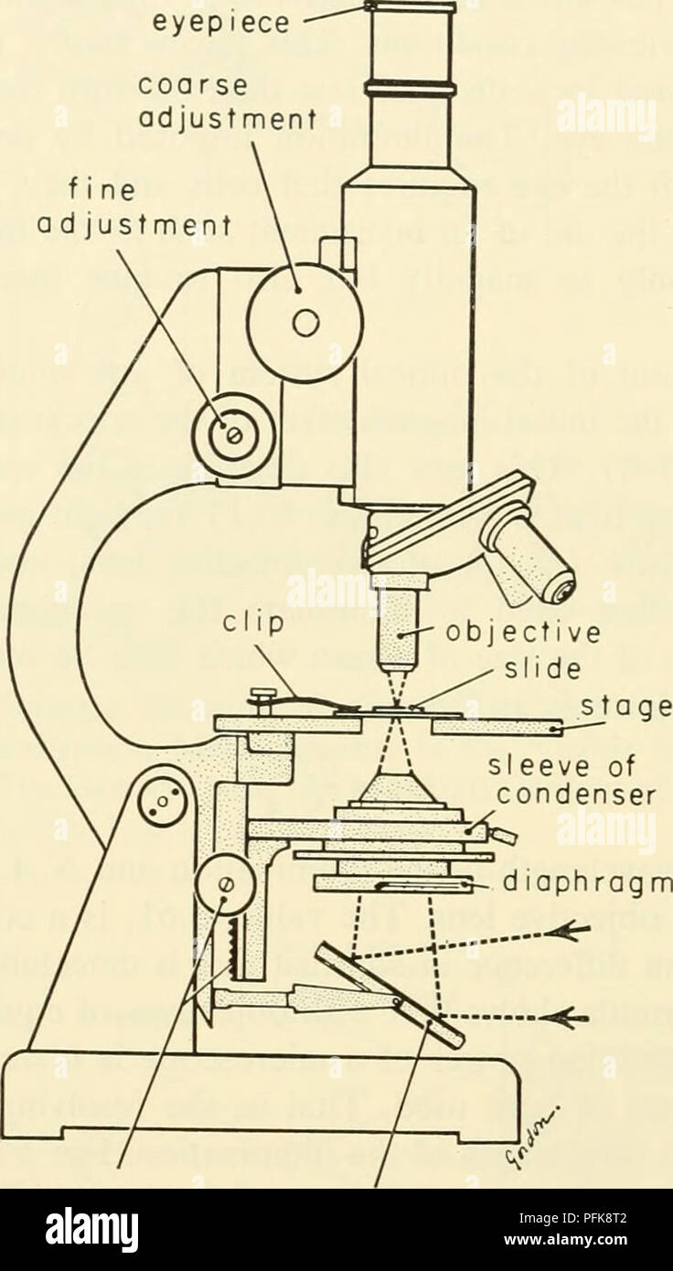
Parts of a Compound Microscope and Their Functions - NotesHippo Compound microscope mechanical parts (Microscope Diagram: 2) include base or foot, pillar, arm, inclination joint, stage, clips, diaphragm, body tube, nose piece, coarse adjustment knob and fine adjustment knob. Base: It's the horseshoe-shaped base structure of microscope. All of the other components of the compound microscope are supported ...

LABELING THE COMPOUND LIGHT MICROSCOPE 2 Diagram | Quizlet
Microscope Parts, Function, & Labeled Diagram - slidingmotion Microscope parts labeled diagram gives us all the information about its parts and their position in the microscope. Microscope Parts Labeled Diagram The principle of the Microscope gives you an exact reason to use it. It works on the 3 principles. Magnification Resolving Power Numerical Aperture. Parts of Microscope Head Base Arm Eyepiece Lens

Simple Microscope - Diagram (Parts labelled), Principle ...
Microscope Parts and Functions With Labeled Diagram and Functions How does a Compound Microscope Work? Before exploring microscope parts and functions, you should probably understand that the compound light microscope is more complicated than just a microscope with more than one lens. Parts of a Chainsaw - Anatomy Explained [2022 Update]

A Study of the Microscope and its Functions With a Labeled ...
Overview of ELISA | Thermo Fisher Scientific - US The antigen is then detected either directly (labeled primary antibody) or indirectly (such as labeled secondary antibody). ... Diagram of common ELISA formats (direct vs. sandwich assays). ... The secondary antibodies are either fluorescent (for direct measurement by a fluorescent plate reader or microscope) or enzyme-conjugated (for detection ...

Parts of a microscope with functions and labeled diagram
Compound Microscope Parts, Functions, and Labeled Diagram Compound Microscope Parts, Functions, and Labeled Diagram Parts of a Compound Microscope Each part of the compound microscope serves its own unique function, with each being important to the function of the scope as a whole.

Compound Microscope- Definition, Labeled Diagram, Principle ...
Binocular Microscope Anatomy - Parts and Functions with a Labeled Diagram Now, I will discuss the details anatomy of the light compound microscope with the labeled diagram. Why it is called binocular: because it has two ocular lenses or an eyepiece on the head that attaches to the objective lens, this ocular lens magnifies the image produced by the objective lens. Binocular microscope parts and functions

This is a common compound microscope. Label its parts from A ...
Parts of a microscope with functions and labeled diagram Sep 17, 2022 · Q. Differentiate between a condenser and an Abbe condenser. Ans. Condensers are lenses that are used to collect and focus light from the illuminator into the specimen. They are found under the stage next to the diaphragm of the microscope. They play a major role in ensuring clear sharp images are produced with a high magnification of 400X and above.

This is a common compound microscope. Label its parts from A ...
Microscope Types (with labeled diagrams) and Functions Compound microscope labeled diagram Compound microscope functions: It finds great application in areas of pathology, pedology, forensics etc Its greater order of magnification allows for deeper study of microbial organisms to Detect the cause of diseases Study the mineral composition in soils
Parts of Stereo Microscope (Dissecting microscope) – labeled ...
Labeled Parts Compound Microscope [4THWBQ] A compound light microscope is a type of light microscope that uses a compound lens system meaning it operates through two sets of lenses to magnify the image of a specimen Two different compound light microscope models with their parts labeled Leica DM1000 Fluorescence Filter - Blue - 11513828 Compound Microscopes Defining Features Image 1 ...

PARTS OF MICROSCOPE| LEARN TO LABEL COMPOUND MICROSCOPE| JUST ...
Label a Compound Microscope Diagram | Quizlet Start studying Label a Compound Microscope. Learn vocabulary, terms, and more with flashcards, games, and other study tools. ... Label this. Illuminator Switch. Sets found in the same folder. A&P II Ch. 24 Digestive Lab QUIZ. 10 terms. CWRN2016. Body planes Label. 9 terms. Hesi_Study.

Compound Microscope Parts – Labeled Diagram and their ...
Labelled Diagram of Compound Microscope The below mentioned article provides a labelled diagram of compound microscope. Part # 1. The Stand: The stand is made up of a heavy foot which carries a curved inclinable limb or arm bearing the body tube. The foot is generally horse shoe-shaped structure (Fig. 2) which rests on table top or any other surface on which the microscope in kept.
Parts Of A Microscope - Parts Of A Compound Microscope, HD ...

Compound Microscope Parts, Functions, and Labeled Diagram ...

Difference between Simple and Compound Microscope ...

16 Basic Parts of Microscope, Function, Names & Labeled Diagram

Compound Microscope Parts – Labeled Diagram and their ...

Compound Microscope: Parts of Compound Microscope

Compound Microscope Parts, Function, & Diagram | What is a ...

Compound Microscope Labeled Diagram | Quizlet

Parts of a Compound Microscope and Their Functions

The Parts of a Compound Microscope and How To Handle Them ...
Parts of a Microscope with Their Functions – Microbe Online
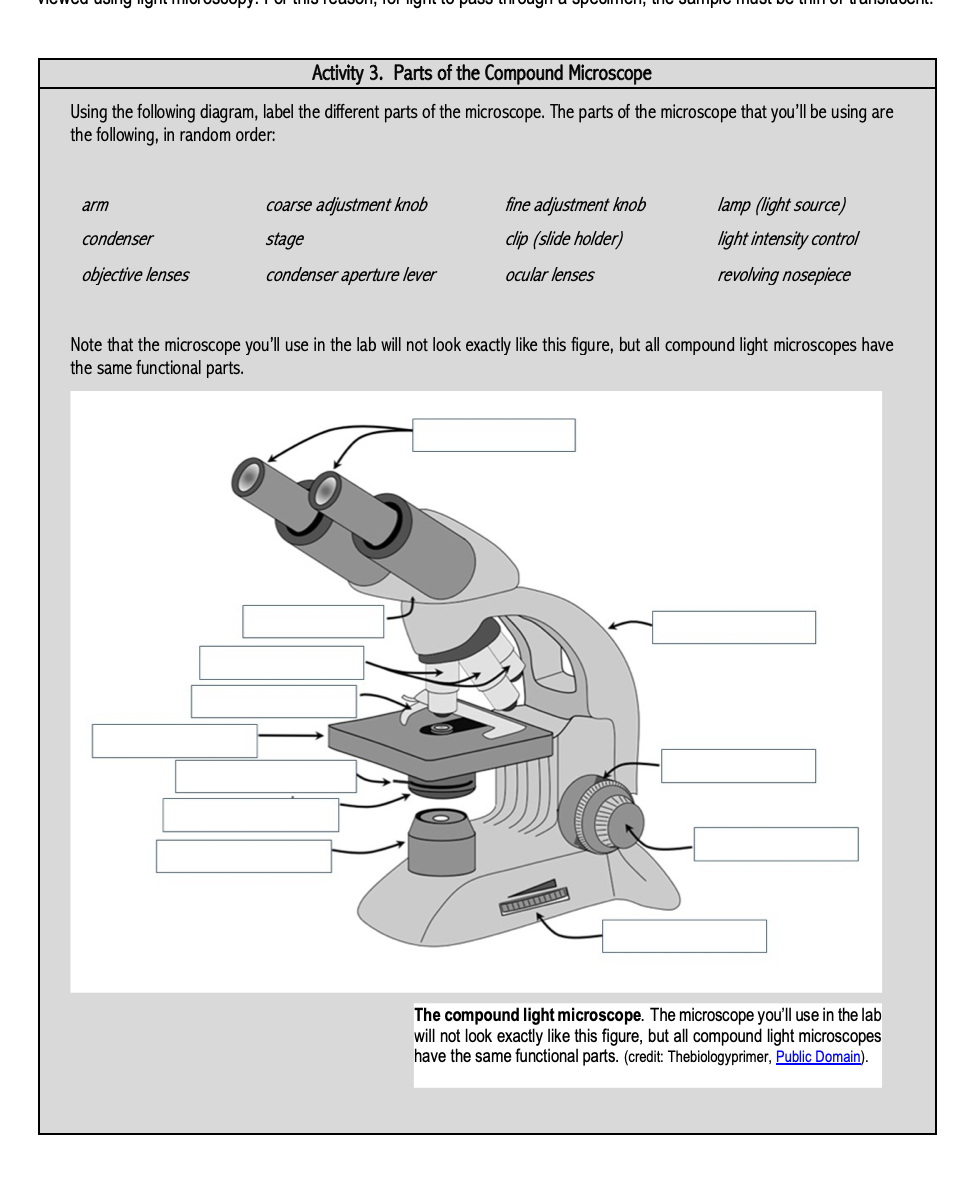
Solved Activity 3. Parts of the Compound Microscope Using ...

Who Invented the Microscope? History of Microscope - Rs ...
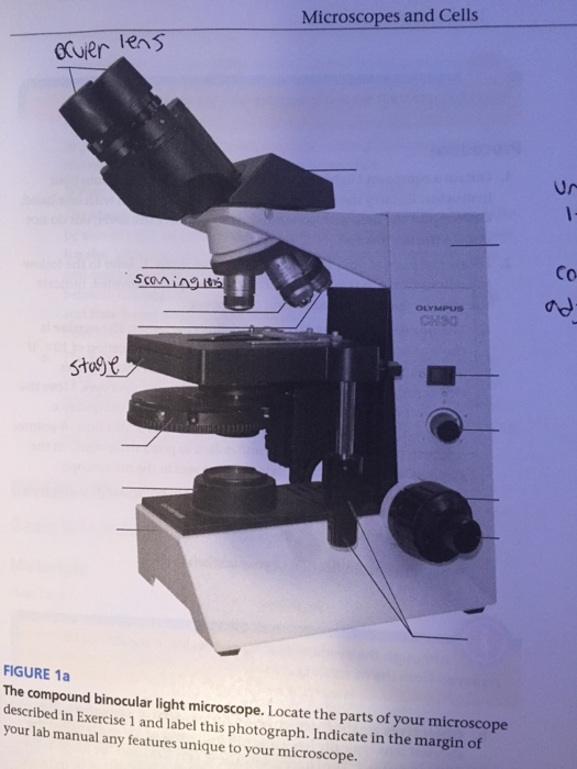
Solved Microscopes and Cells Gruer Co FIGURE 1a The compound ...

Microscope Labeling

Parts of a Microscope - SmartSchool Systems
Draw a neat labeled ray diagram of a compound microscope ...
16 Parts of a Compound Microscope: Diagrams and Video ...
Parts of a microscope with functions and labeled diagram

Simple Microscope Definition, Magnification, Parts And Uses

Compound Microscope Parts, Functions, and Labeled Diagram ...

General Biology | Carlson Stock Art | General biology ...

Diagram of a Compound Microscope

Compound Microscope – Diagram (Parts labelled), Principle and ...
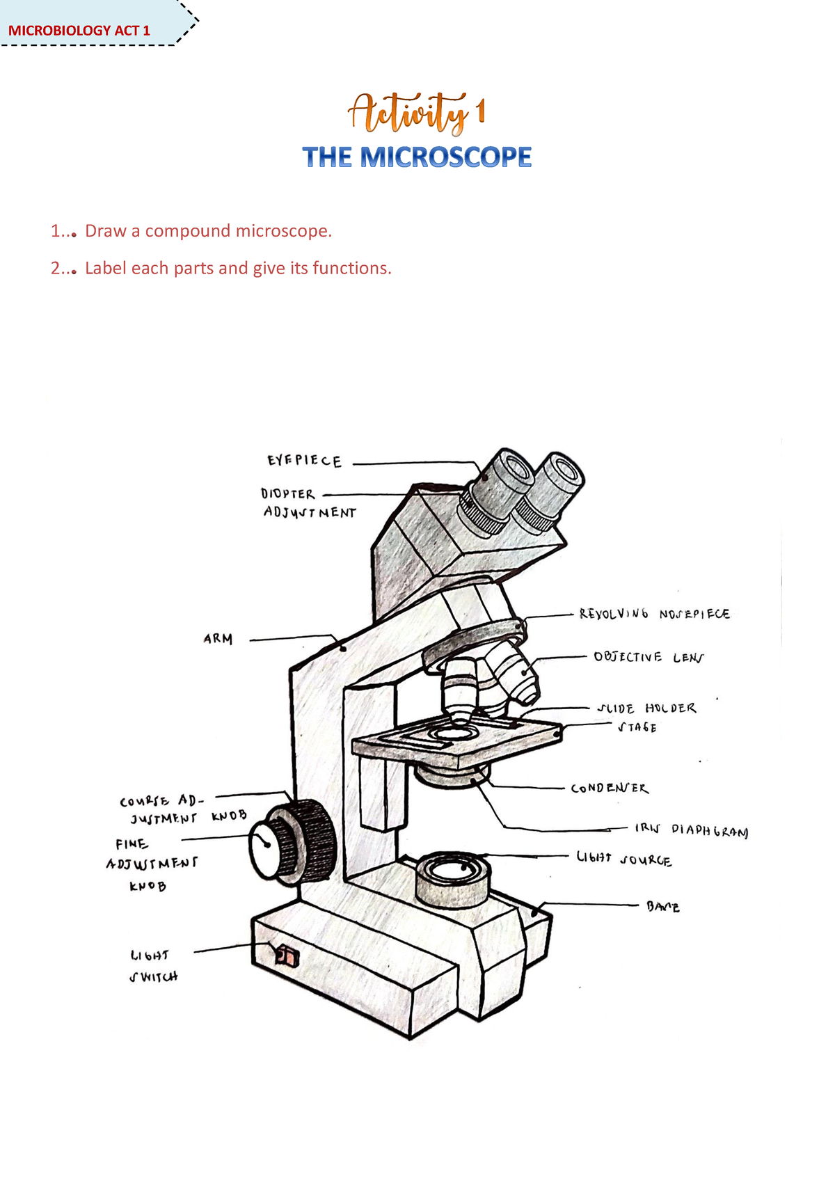
Microscope Activity - MICROBIOLOGY - 1... Draw a compound ...
MICROBIO 16 Parts of a Compound Microscope with Diagram and ...

Solved A. OLYMPUS C. B. Use the Diagram to answer the | Chegg.com
Cytology. Cytology. radiation used to illuminate the specimen ...
Parts of a microscope with functions and labeled diagram

Compound microscope - their parts and function - Microscopy4kids
Post a Comment for "45 compound microscope diagram labeled"