45 diagram for labelling microscope
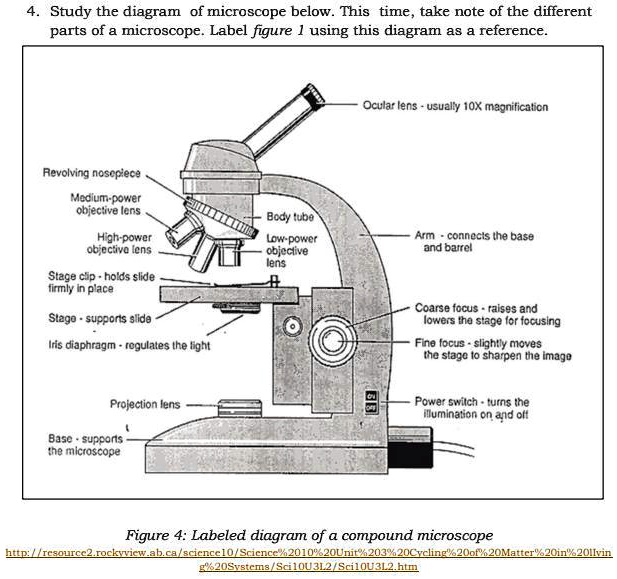
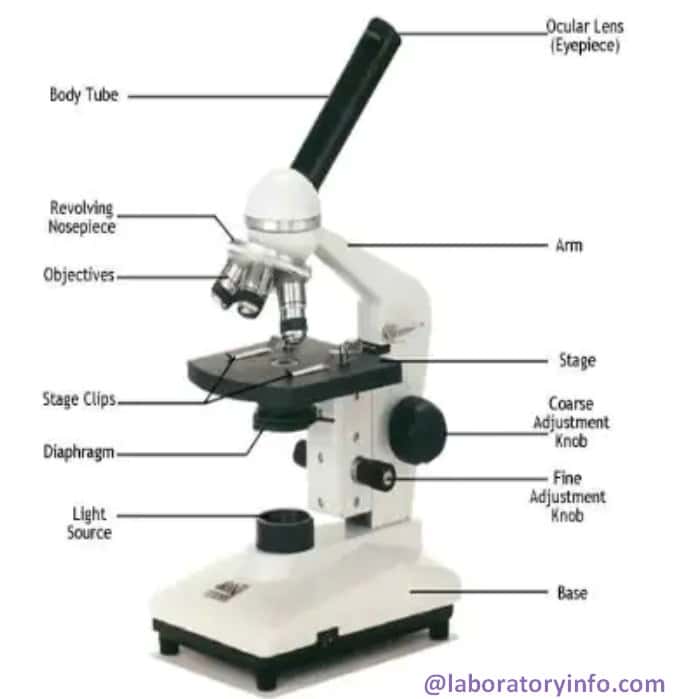
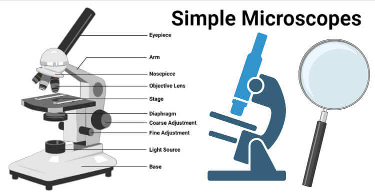
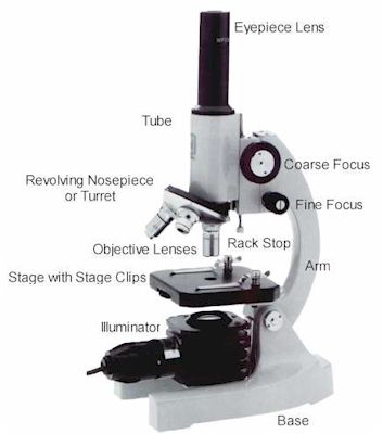
Microscope Labeling Diagram | Quizlet Microscope Labeling 4.2 (66 reviews) Flashcards Learn Test Match Created by PGFry210 Teacher Terms in this set (12) Term Body Tube Definition Separates the eyepiece lens from the objective lenses. Location Term Eyepiece Definition Contains the ocular lens and magnifies the image produced by the objective lenses. Location Term Coarse Focus Knob Parts of the Microscope (Labeled Diagrams) Simple microscope labelled diagram Image created with Biorender Tube/Body Tube It serves as the connector between the eyepiece/ocular and objective lenses. Objective lenses The lenses have varying magnifying power, which typically consists of 10x, 40x, and 100x.
Compound Microscope: Definition, Diagram, Parts, Uses, Working Principle A compound microscope is defined as. A microscope with a high resolution and uses two sets of lenses providing a 2-dimensional image of the sample. The term compound refers to the usage of more than one lens in the microscope. Also, the compound microscope is one of the types of optical microscopes. The other type of optical microscope is a ...
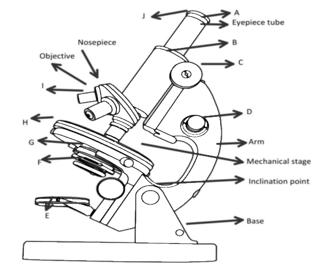
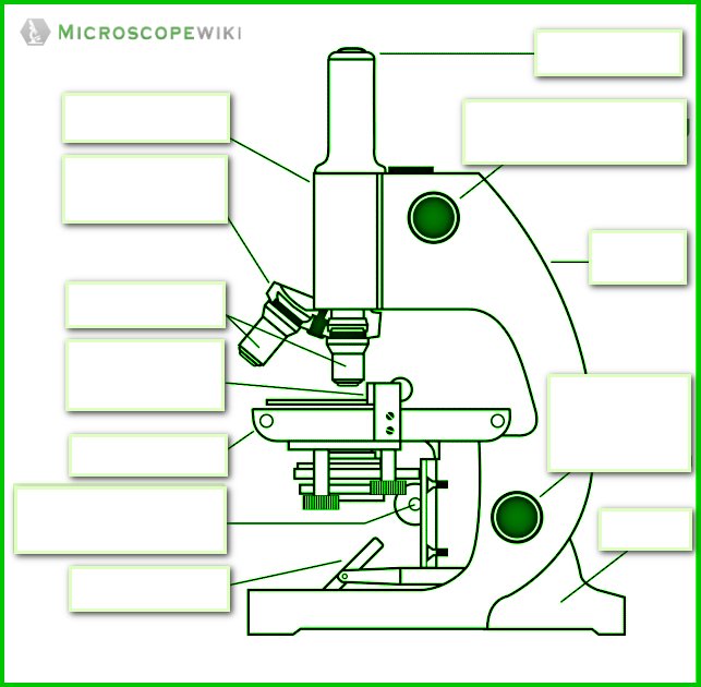
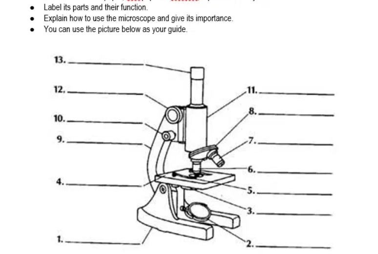
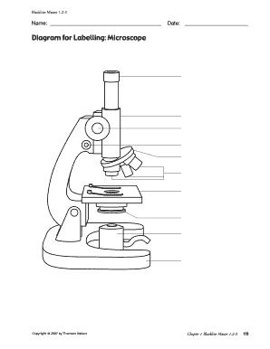
Diagram for labelling microscope
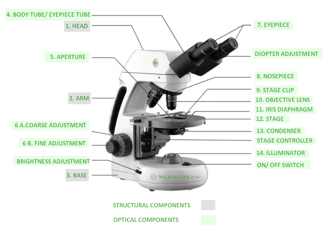
Parts of a microscope with functions and labeled diagram - Microbe Notes Figure: Diagram of parts of a microscope There are three structural parts of the microscope i.e. head, base, and arm. Head - This is also known as the body. It carries the optical parts in the upper part of the microscope. Base - It acts as microscopes support. It also carries microscopic illuminators. Label a microscope - Teaching resources Y6 Labelling Microscope Labelled diagram. by Sciencedept. KS2 Y6 Science. Electron Microscope Group sort. by Msxmdg. KS3 KS4 Science. French Label a Compass (8 points) Labelled diagram. by Kgandrew87. French. Label a Human Skeleton Labelled diagram. by Harrisond. KS1 KS2 Y2 Y3 Y4 Y5 Y6 Science. Labelling A Microscope Teaching Resources | Teachers Pay Teachers Print & Go Notes - No Prep Needed!This resource includes student notes, a teacher key, and slides for teaching. It follows the Alberta Science 10 Unit C (Biology) Curriculum.Student Notes (7 pages) covers the following topics:microscope diagram - labelling parts and their functionstimeline of the development of microscopes & cell theorycalculations (magnification, field of view, actual size ...
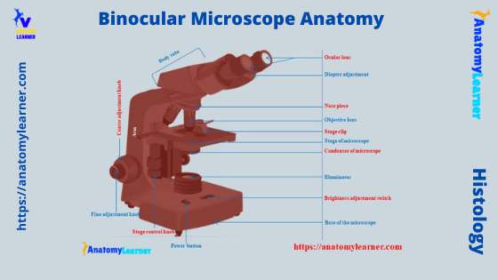
Diagram for labelling microscope. Compound Microscope Parts, Functions, and Labeled Diagram Meiji MT-30 Binocular Microscope - Rechargeable. $618.55. Labomed 9135010 CxL Binocular Cordless Microscope, 4x, 10x, 40x Objectives, LED Illumination. $713.21. ACCU-SCOPE EXM-150-MS Monocular Cordless Microscope with Mechanical Stage, Rechargeable. $325.00. Get relevant offers, the latest promotions, and articles from New York Microscope Company. PDF Label parts of the Microscope: Answers Label parts of the Microscope: Answers Coarse Focus Fine Focus Eyepiece Arm Rack Stop Stage Clip . Created Date: 20150715115425Z ... Label the microscope — Science Learning Hub Use this interactive to identify and label the main parts of a microscope. Drag and drop the text labels onto the microscope diagram. diaphragm or iris base eye piece lens fine focus adjustment light source stage coarse focus adjustment high-power objective Download Exercise Labeling the Parts of the Microscope | Microscope World Resources This activity has been designed for use in homes and schools. Each microscope layout (both blank and the version with answers) are available as PDF downloads. You can view a more in-depth review of each part of the microscope here. Download the Label the Parts of the Microscope PDF printable version here.
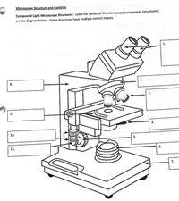
Labelling a Microscope Diagram | Quizlet The function of the microscope stage is to allow for easy movement and manipulation of the slide. This will allow you to focus on the specimen in an accurate manner. What is the diaphragm? A diaphragm on a microscope is the piece that enables the user to adjust the amount of light that is focused under the specimen being observed. Light Source. A Study of the Microscope and its Functions With a Labeled Diagram ... These labeled microscope diagrams and the functions of its various parts, attempt to simplify the microscope for you. However, as the saying goes, 'practice makes perfect', here is a blank compound microscope diagram and blank electron microscope diagram to label. Microscope Labeling Practice Diagram | Quizlet Put your eye here. Contains one lens of 10 power magnification. Arm Supports the body tube and stage. Carry with one hand here. Stage Where the microscope slide is placed for viewing. Coarse Focus Knob (Coarse Adjustment) Elevates or lowers the stage a large distance with each turn of the knob. Fine Focus Knob (Fine Adjustment) PDF Label parts of the Microscope Label parts of the Microscope: . Created Date: 20150715115425Z
Microscope Labeling - The Biology Corner The labeling worksheet could be used as a quiz or as part of direct instruction where students label the microscope as you go over what each part is used for. The google slides shown below have the same microscope image with the labels for students to copy. Compound Microscope - Diagram (Parts labelled), Principle and Uses A compound microscope: Is used to view samples that are not visible to the naked eye. Uses two types of lenses - Objective and ocular lenses. Has a higher level of magnification - Typically up to 2000x. Is used in hospitals and forensic labs by scientists, biologists and researchers to study microorganisms. Invented in the late 16th century ... Microscope Parts and Functions Microscope Parts and Functions With Labeled Diagram and Functions How does a Compound Microscope Work? Before exploring microscope parts and functions, you should probably understand that the compound light microscope is more complicated than just a microscope with more than one lens. Microscopy: Intro to microscopes & how they work (article) - Khan Academy In scanning electron microscopy ( SEM ), a beam of electrons moves back and forth across the surface of a cell or tissue, creating a detailed image of the 3D surface. This type of microscopy was used to take the image of the Salmonella bacteria shown at right, above.
Simple Microscope - Diagram (Parts labelled), Principle, Formula and Uses A simple microscope consists of Optical parts Mechanical parts Labeled Diagram of simple microscope parts Optical parts The optical parts of a simple microscope include Lens Mirror Eyepiece Lens A simple microscope uses biconvex lens to magnify the image of a specimen under focus.
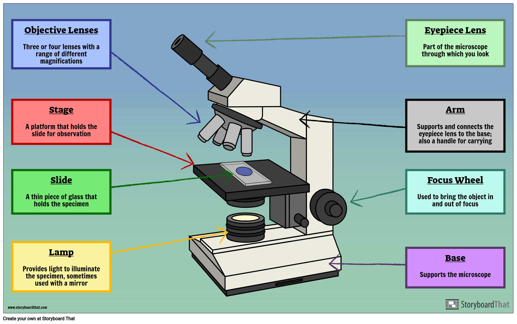
Label a Microscope - Storyboard That Create a poster that labels the parts of a microscope and includes descriptions of what each part does. Click "Start Assignment". Use a landscape poster layout (large or small). Search for a diagram of a microscope. Using arrows and textables label each part of the microscope and describe its function. Copy This Storyboard.
Labelling A Microscope Teaching Resources | Teachers Pay Teachers Print & Go Notes - No Prep Needed!This resource includes student notes, a teacher key, and slides for teaching. It follows the Alberta Science 10 Unit C (Biology) Curriculum.Student Notes (7 pages) covers the following topics:microscope diagram - labelling parts and their functionstimeline of the development of microscopes & cell theorycalculations (magnification, field of view, actual size ...
Label a microscope - Teaching resources Y6 Labelling Microscope Labelled diagram. by Sciencedept. KS2 Y6 Science. Electron Microscope Group sort. by Msxmdg. KS3 KS4 Science. French Label a Compass (8 points) Labelled diagram. by Kgandrew87. French. Label a Human Skeleton Labelled diagram. by Harrisond. KS1 KS2 Y2 Y3 Y4 Y5 Y6 Science.
Parts of a microscope with functions and labeled diagram - Microbe Notes Figure: Diagram of parts of a microscope There are three structural parts of the microscope i.e. head, base, and arm. Head - This is also known as the body. It carries the optical parts in the upper part of the microscope. Base - It acts as microscopes support. It also carries microscopic illuminators.

















Post a Comment for "45 diagram for labelling microscope"