41 label the photomicrogram of the trachea
(Get Answer) - Determine the angle of i, r and q this is reflection of ... Determine the angle of i, r and q this is reflection of light General Structure of Mucosa Label the structures that comprise the respiratory tract mucosa (mucous membrane). ... lung Segmental bronchus Trachea prey="" 11="" of="" 46="" next=""> Trachea histology of respiratory system low power Label the photomicrogram of the trachea. A&P 139 Chapter 19 Flashcards | Quizlet Label the photomicrogram of the lung. Label the photomicrogram of the trachea. Cricoid. Which of these laryngeal cartilages is single? Label these structures of the upper respiratory system. tidal volume. The volume of air that enters (or leaves) during a single respiratory cycle is the.
A&P 2 Lab Practical Final Flashcards - Quizlet Larynx Label the photomicrogram of the lung. Put the following structures of the lower respiratory tract in order from proximal to distal. Label these structures of the upper respiratory system. Label the anterior view of the larynx based on the hints if provided.

Label the photomicrogram of the trachea
The Bronchi: Anatomy, Function, and Treatment - Verywell Health The bronchi are the airways that lead from the trachea into the lungs and then branch off into progressively smaller structures until they reach the alveoli, the tiny sacs that allow for the exchange of oxygen and carbon dioxide in the lungs. While the bronchi function primarily as passageways for air, they also play a role in immune function. Free Automated Malware Analysis Service - powered by Falcon Sandbox ... Submit malware for free analysis with Falcon Sandbox and Hybrid Analysis technology. Hybrid Analysis develops and licenses analysis tools to fight malware. Labeled diagram of the lungs/respiratory system. - SERC View Original Image at Full Size. Labeled diagram of the lungs/respiratory system. Image 37789 is a 1125 by 1408 pixel PNG Uploaded: Jan10 14. Last Modified: 2014-01-10 12:15:34
Label the photomicrogram of the trachea. A & P lab test 4 Flashcards - Quizlet Select all that apply. Label these structures of the upper respiratory system. Correctly label the components of the upper respiratory tract. Label the anterior view of the lower respiratory tract based on the hints if provided. Correctly label the components of the lungs. Correctly label the components of the pulmonary alveoli. BIO208 Lab Practical 2 - 10/6/2019 Lab Practical 2 Home - Course Hero 10/6/2019 Lab Practical 2 Question Label the structure with a "star" symbol beside it. 4 Incorrect Mark 0.00 out of 1.00 Answer: trachea The correct answer is: larynx (based on the document attached to the question as reference) A&P 2 Lab Respiratory System Flashcards - Quizlet Label the lateral view of the larynx based on the hints if provided. Image: Label the lateral view of ... Image: Label the photomicrogram of the trachea. The Trachea (Human Anatomy): Picture, Function, Conditions, and More The trachea, commonly known as the windpipe, is a tube about 4 inches long and less than an inch in diameter in most people. The trachea begins just under the larynx (voice box) and runs down...
Histology of trachea and lung - SlideShare 1. HISTOLOGY OF TRACHEA AND LUNG Dr.ushakannan,Asst.professor. 2. RESPIRATORY SYSTEM Conducting Part- responsible for passage of air and conditioning of the inspired air. Examples:nasal cavities,pharynx, trachea, bronchi and their intrapulmonary continuations. Respiratory Part-involved with the exchange of oxygen and carbondioxide between blood ... Can you label the lungs? Quiz - PurposeGames.com This is an online quiz called Can you label the lungs? There is a printable worksheet available for download here so you can take the quiz with pen and paper. From the quiz author. Labeling the lungs. This quiz has tags. Click on the tags below to find other quizzes on the same subject. lungs. respiratory system. A&P 139 Chapter 19 Flashcards | Quizlet Label the photomicrogram of the lung. Label the photomicrogram of the trachea. Cricoid. Which of these laryngeal cartilages is single? Label these structures of the upper respiratory system. tidal volume. The volume of air that enters (or … A&P 2 Lab Unit 2 Flashcards - Quizlet Label the lateral view of the larynx based on the hints if provided. Image: Label the lateral view of ... Image: Label the photomicrogram of the trachea.
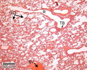
Label The Photomicrograph Of The Lung : 4 Chloro Dl Phenylalanine ... The lower respiratory system., put the following layers of the trachea in order from superficial to deep., label the structures of the upper respiratory . Label the photomicrogram of the lung segmental branch of pulmonary a. Relative amounts of glands, cartilage, smooth muscles and connective tissue fibers present in the wall of the tubes. Anatomy A215 Virtual Microscopy - Indiana University Bloomington Anatomy A215 Virtual Microscopy. Each alveolus is a small air space surrounded by an extensive capillary network. The epithelium which lines the alveoli is an extremely thin simple squamous in close proximity to the. capillary walls. Alveoli make up the major part of the lung. A&P 2 Lab Unit 2 Flashcards - Quizlet Label the lateral view of the larynx based on the hints if provided. Image: Label the lateral view of ... Image: Label the photomicrogram of the trachea. A&P 2 Lab Unit 2 Flashcards | Quizlet Label the photomicrogram of the trachea. Identify these structures in the left-sided midsagittal view of the superior portion of the lower respiratory system. Identify the anatomical structures shown in the picture of the thorax. Identify the anatomical structures shown in a lateral view of the left lung.
Respiratory Epithelium: Trachea
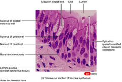
Lab 2: Microscopy and the Study of Tissues - UW-La Crosse The lining of the trachea consists of a type of tissue called pseudostratified (ciliated) columnar epithelium. This single layer of ciliated cells appears stratified because the cells vary in their thickness and because their nuclei are located at different levels. 2 - Pseudostratified columnar epithelium (close-up view) Ciliated border

Respiratory Epithelium Pictures Flashcards | Quizlet
Histology, Alveolar Cells - StatPearls - NCBI Bookshelf Alveoli represent the major sites of gas exchange. Their presence increases the surface area of the lung to maximize gas exchange, much like villi and microvilli increase the absorptive surface area of the digestive tract. For alveoli to carry out their function efficiently, they must be both a dynamic and stable system. The lung parenchyma must be able to expand and recoil during inspiration ...

Photomicrograph Of Pseudostratified Ciliated Columnar Epithelium Of The ...
A&P 2 Lab Unit 2 Flashcards - Quizlet Label the lateral view of the larynx based on the hints if provided. Image: Label the lateral view of ... Image: Label the photomicrogram of the trachea.

ENT for medical students: TRACHEA - Anatomy
Solved Label the photomicrogram of the lung Segmental branch | Chegg.com Answer to Solved Label the photomicrogram of the lung Segmental branch
Anatomy Lab Exam 1 Flashcards | Easy Notecards
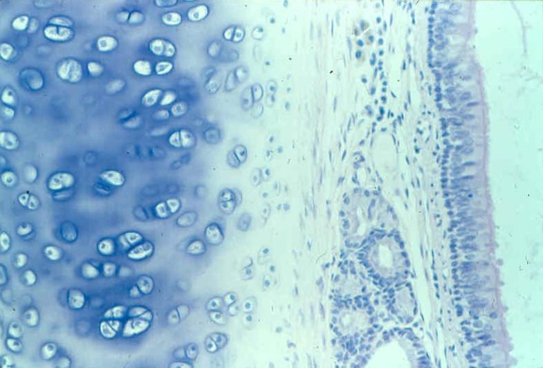
Solved Label the photomicrogram of the trachea. Cilia Lamina | Chegg.com Anatomy and Physiology questions and answers. Label the photomicrogram of the trachea. Cilia Lamina propria Submucosa Cilia Basement membrane Submucosa Epithelium Basement membrane Lamina propria Epithellum. Question: Label the photomicrogram of the trachea. Cilia Lamina propria Submucosa Cilia Basement membrane Submucosa Epithelium Basement ...

Trachea, Anatomy, Respiratory
A & P lab test 4 Flashcards | Quizlet Label the photomicrogram of the trachea. Correctly label the following anatomical features of the lower respiratory tract. ... Drag each label into the appropriate position in order to identify whether the term or item is involved with chemical or mechanical digestion.
Respiratory: The Histology Guide
A&P 2 lab ex 33 Flashcards - Quizlet Label the lateral view of the larynx based on the hints if provided. Image: Label the lateral view of ... Image: Label the photomicrogram of the trachea.

Anatomy And Physiology Archive | October 25, 2017 | Chegg.com
Solved General Structure of Mucosa Label the structures that | Chegg.com This problem has been solved! See the answer Show transcribed image text Expert Answer 100% (1 rating) 1.UPPER LOBE OF RIGHT LUNG 2.RIGHT MAIN BRONCHUS 3. SEGMENTAL BRONCHUS 4. TRACHEA 5. UPPER LOBE OF LEFT LUNG 6.LEFT MAIN BRONCHUS 1.MUCUS 2.GOBL … View the full answer

Pin on Voice and related anatomy/physiology
M4 Respiratory system Flashcards | Quizlet visual examination of the bronchi using an endoscope inserted through the mouth and trachea for direct viewing of structures or for projection on a monitor. laryngoscopy. visual examination of the larynx to detect tumors, foreign bodies, nerve or structural injury, or other abnormalities.
Respiratory: The Histology Guide
Anatomy diagrams week 1-2 Flashcards - Quizlet Label the lateral view of the larynx based on the hints if provided. Image: Label the lateral view of ... Image: Label the photomicrogram of the trachea.
The respiratory mucosa lines the conduction portion the air ...
Trinidad State College Home Page Trinidad State is a Hispanic-Serving Institution (HSI) HSI is defined in federal law (the Higher Education Opportunity Act, Title V, 2008) as an accredited, degree-granting, public or private nonprofit institution of higher education with 25% or more total undergraduate Hispanic full-time equivalent (FTE) student enrollment.

110 best Histology - Respiratory images on Pinterest | Lunges, Lungs ...
Labeled diagram of the lungs/respiratory system. - SERC View Original Image at Full Size. Labeled diagram of the lungs/respiratory system. Image 37789 is a 1125 by 1408 pixel PNG Uploaded: Jan10 14. Last Modified: 2014-01-10 12:15:34

Respiratory Epithelium Pictures Flashcards | Quizlet
Free Automated Malware Analysis Service - powered by Falcon Sandbox ... Submit malware for free analysis with Falcon Sandbox and Hybrid Analysis technology. Hybrid Analysis develops and licenses analysis tools to fight malware.
![New Page 1 [www.galeps.org]](http://www.galeps.org/jadams/biol 2213/Images/Respiratory/Unmodified/Best trachea section.jpg)
New Page 1 [www.galeps.org]
The Bronchi: Anatomy, Function, and Treatment - Verywell Health The bronchi are the airways that lead from the trachea into the lungs and then branch off into progressively smaller structures until they reach the alveoli, the tiny sacs that allow for the exchange of oxygen and carbon dioxide in the lungs. While the bronchi function primarily as passageways for air, they also play a role in immune function.

CLASS BLOG
Post a Comment for "41 label the photomicrogram of the trachea"