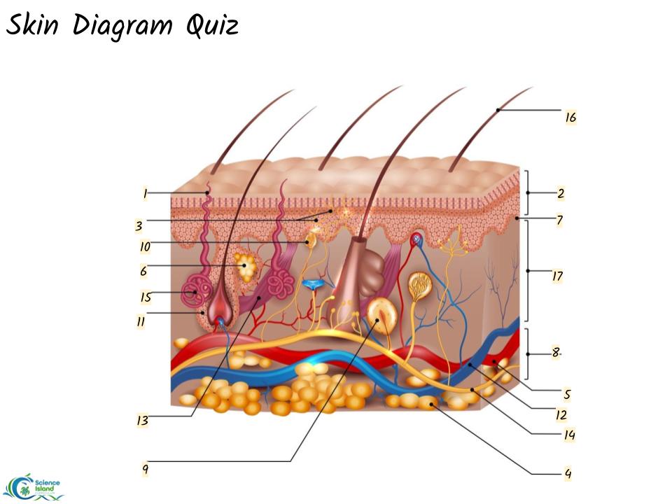
40 skin diagram to label
Anatomy, Skin (Integument), Epidermis - StatPearls - NCBI Bookshelf Skin is the largest organ in the body and covers the body's entire external surface. It is made up of three layers, the epidermis, dermis, and the hypodermis, all three of which vary significantly in their anatomy and function. The skin's structure is made up of an intricate network which serves as the body's initial barrier against pathogens, UV light, and chemicals, and mechanical injury. 6,243 Skin Diagram Stock Photos and Images - 123RF 6,243 skin diagram royalty-free stock photos and images found for you. Page of 63 Wound healing. stages of the post-trauma repairing process. hemostasis, inflammatory, proliferative, and remodeling phase. cross section of a layers of the human skin Dry and hydrated skin layer with ceramide and woman illustration. beauty and skin care concept
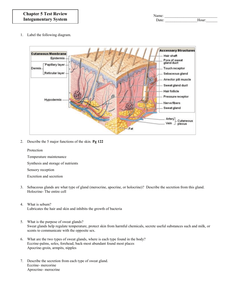
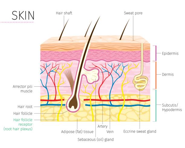
5.1 Layers of the Skin - Anatomy and Physiology 2e - OpenStax Stratum Basale. The stratum basale (also called the stratum germinativum) is the deepest epidermal layer and attaches the epidermis to the basal lamina, below which lie the layers of the dermis. The cells in the stratum basale bond to the dermis via intertwining collagen fibers, referred to as the basement membrane. A finger-like projection, or fold, known as the dermal papilla (plural ...

Skin diagram to label
Anatomy of the Skin - Stanford Children's Health The skin is made up of 3 layers. Each layer has certain functions: Epidermis. Dermis. Subcutaneous fat layer (hypodermis) ... Anatomy of the Epidermis with Pictures - Verywell Health The epidermis is composed of layers of skin cells called keratinocytes. Your skin has four layers of skin cells in the epidermis and an additional fifth layer in areas of thick skin. The four layers of cells, beginning at the bottom, are the stratum basale, stratum spinosum, stratum granulosum, and stratum corneum. Skin structure diagram - Teaching resources - Wordwall L3 skin diagram NEW Labelled diagram by Kmiller14 Skin structure Match up by Siobhan45 Skin structure Match up by Kellyrandall Level 3 Advanced Skin Diagram Labelled diagram by Lauragalgey Beauty Therapy Skin diagram to label Labelled diagram by Wyeoman Technical education Biology Skin Diagram (L3 camouflage) Labelled diagram by Kmiller14
Skin diagram to label. 37 Labeled Skin Diagram Illustrations & Clip Art - iStock Labeled Skin Diagram stock illustrations View labeled skin diagram videos Browse 37 labeled skin diagram stock illustrations and vector graphics available royalty-free, or start a new search to explore more great stock images and vector art. Sort by: Most popular Systemic lupus erythematosu Systemic lupus erythematosu. Skin Diagram || How to draw and label the parts of skin 'Skin Diagram || How to draw and label the parts of skin' is demonstrated in this video tutorial step by step.The sense of touch had received supreme importa... Structure of the Skin: Cross-section through the Skin, Diagrams The skin comprises three different layers: The outermost layer: Epidermis The middle layer: Dermis The innermost layer: Hypodermis Learn About Skin and Its Functions Fig: The Cross-Section Through the Skin Receptors in the Skin Skin serves as a sense organ and is also the largest sense organ. It possesses a number of sensory receptors. skin labeling Diagram | Quizlet Middle layer of the skin; contains collagen; location of sweat & sebaceous glands and nerve endings and capilaries Epidermis outermost layer of the skin;composed of squamous epithelium; contains keratin subcutaneous layer Innermost layer of skin, contains fat and is the location of main blood vessels basal layer of epidermis
Skin diagram - Teaching resources - Wordwall The Skin Labelled diagram by Djspencer1 University skin Match up by 22098368 Skin Random wheel by Gailwoodward skin Match up by Annerollins Break Even Diagram Labelled diagram by Tiles KS4 KS5 Y10 Y11 Y12 Break Even Business Studies Finance Finances Fixed Costs OCR GCSE Business Variable Costs Harry Potter (diagram) Labelled diagram Skin Layers: Structure, Function, Anatomy, and More - Verywell Health There are three main layers of skin: Epidermis: The outermost layer, which contains five sub-layers Dermis: The middle layer, which consists of two parts known as the papillary dermis (thin, upper layer) and the reticular dermis (thick, lower layer) Subcutaneous tissue: The deepest layer of skin What is the integumentary system? A Human Body Skin-structure Quiz! - ProProfs Quiz Skin Structure C. Root 2. Label A is: A. Hair Root B. Hair Follicle Receptor C. Hair Shaft D. Hair Follicle 3. Label B is: A. Meissner's Corpuscles B. Pacinian Corpuscle C. Pore D. Free Nerve Ending 4. Label C is: A. Dermal Papillae Layer B. Arrector Pili Muscle C. Sebaceous (oil) Gland D. Artery 5. Label D is: A. Sebaceous (oil) gland B. Label the Skin Quiz - PurposeGames.com Label the Skin by LegoA1 86,589 plays 11 questions ~30 sec English 45 4.36 (you: not rated) Tries Unlimited [?] Last Played February 22, 2022 - 12:00 am There is a printable worksheet available for download here so you can take the quiz with pen and paper. Remaining 0 Correct 0 Wrong 0 Press play! 0% 0:00.0 Highscores Show More
Skin Diagram - Etsy Integumentary Anatomy Coloring Page- LABELED- Digital Download Skin Anatomy Diagram Anatomy Worksheet Med Student Study Guide Anatomy Art JennMedArt (66) $3.90 Vintage 1907 German Medical Anatomy Diagram Bookplate Skin Disease Syphilis Spots Pustules Thepapermuseum (351) $23.20 FREE shipping More colors The Skin (Human Anatomy): Picture, Definition, Function, and Skin ... The skin is the largest organ of the body, with a total area of about 20 square feet. The skin protects us from microbes and the elements, helps regulate body temperature, and permits the... A diagrammatic representation of the structure of human skin in cross... A diagrammatic representation of the structure of human skin in cross section. The epidermis is composed of the stratum corneum and the viable epidermis. Layers of the Skin | Anatomy and Physiology I - Lumen Learning The skin is composed of two main layers: the epidermis, made of closely packed epithelial cells, and the dermis, made of dense, irregular connective tissue that houses blood vessels, hair follicles, sweat glands, and other structures. Beneath the dermis lies the hypodermis, which is composed mainly of loose connective and fatty tissues.
Skin Diagram || How to draw and label the parts of skin ...
Integumentary system parts: Quizzes and diagrams | Kenhub Labeled diagram of the skin So what's the idea? Spend some time analyzing the skin diagram labeled above. Try to memorize the appearance and location of each structure. Learning the function of each structure will accelerate your ability to memorize, so be sure to check out our detailed article on The Integumentary System parts and functions .
Solved] I. Parts of the skin and meaning... | Course Hero
Layers of Skin: How Many, Diagram, Model, Anatomy, In Order The epidermis is the top layer of your skin. It's the only layer that is visible to the eyes. The epidermis is thicker than you might expect and has five sublayers. Your epidermis is constantly...
Label a diagram of the skin - Mrs. Sanborn's Science Class
The Skin - Science Quiz - GeoGuessr The Skin - Science Quiz: The skin is an organ. In fact, it's the body's largest organ and is responsible for protection against germs that can cause infection. It also gives us our sense of touch and helps control the body's temperature. The skin is an amazing part of the human body, and this science quiz game will help you identify its components. The middle section of the skin is ...
top_mob16154556682046676786ans ...
Human Skin Diagram & Function | How Many Layers of Skin Are There ... There are a total of 7 layers to the skin. These layers are as follows: Stratum corneum Stratum lucidum Stratum granulosum Stratum spinosum Stratum basale Papillary Layer Reticular Layer Although...
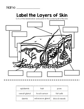
Label the Layers of Skin by Little Learning Lane | TPT
Skin Labeling Quiz - PurposeGames.com Skin Labeling by marthamae 180,968 plays 17 questions ~40 sec English 66 4.12 (you: not rated) Tries Unlimited [?] Last Played January 18, 2023 - 04:38 PM There is a printable worksheet available for download here so you can take the quiz with pen and paper. From the quiz author Epidermis, Dermis, Hypodermis Remaining 0 Correct 0 Wrong 0

A Human Body Skin-structure Quiz! - ProProfs Quiz
Skin Diagram To Label - Simple - cullenpic07.blogspot.com Labeled medical diagram, a 3d cross section of human skin layers and parts such as a hair follicle and sweat glands on a white background. This diagram had been modified from . (0.5 each) oil gland growth layer sweat gland dermis dead cells sebum (oil) hair follicle blood vessels hypodermis epidermis pore nerve the skin the skin .
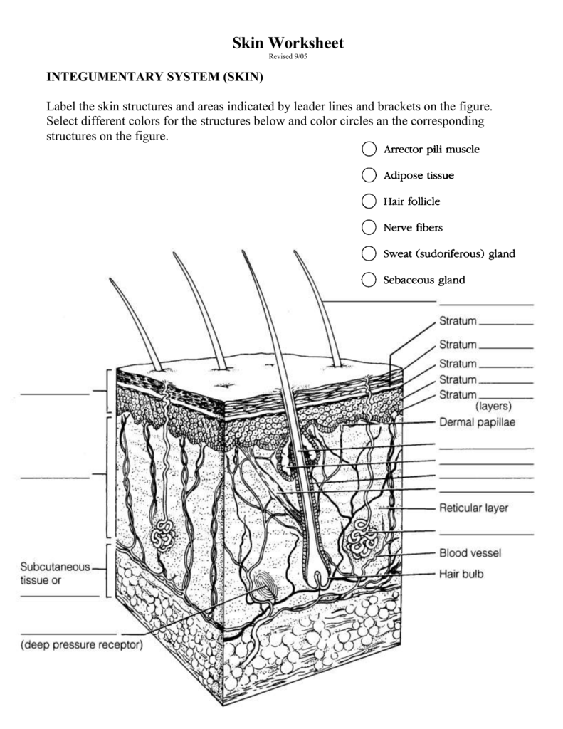
Skin Worksheet
Skin Structure labeling Diagram | Quizlet Start studying Skin Structure labeling. Learn vocabulary, terms, and more with flashcards, games, and other study tools.
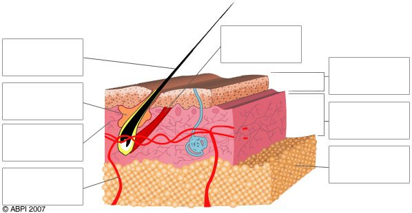
Quiz - Skin - ABPI - Resources for Schools
Labeled Skin Diagram Pictures, Images and Stock Photos Labeled medical diagram, a 3D cross section of human skin layers and parts such as a hair follicle and sweat glands on a white background. Oily & dry skin. Oily & dry skin. Different. Human Skin types and conditions. A diagrammatic sectional view of the skin. Skin layers with glands (sebaceous and sweat glands).
Label the skin - Teaching resources
5.1 Layers of the Skin - Anatomy & Physiology Skin that has four layers of cells is referred to as "thin skin." From deep to superficial, these layers are the stratum basale, stratum spinosum, stratum granulosum, and stratum corneum. Most of the skin can be classified as thin skin. "Thick skin" is found only on the palms of the hands and the soles of the feet.
Anatomy of the Skin
Skin Diagram with Detailed Illustrations and Clear Labels - BYJUS Skin Diagram Skin Diagram The largest organ in the human body is the skin, covering a total area of about 1.8 square meters. The skin is tasked with protecting our body from external elements as well as microbes. Interesting Note:
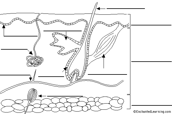
Label Skin Diagram Printout - EnchantedLearning.com
Skin diagram labeled - Healthiack Skin diagram labeled This brief article displays Skin diagram labeled … Please click on the diagram (s) to view larger version. You are welcome to browse healthiack.com for more details on this very topic. Best viewed on 1280 x 768 px resolution in any modern browser. Skin diagram labeled 1075 Skin diagram labeled 1077 Skin diagram labeled 1080

6.5: Laboratory Activities and Assignment - Biology LibreTexts
109 Skin Diagram To Label Illustrations & Clip Art - iStock Choose from 109 Skin Diagram To Label stock illustrations from iStock. Find high-quality royalty-free vector images that you won't find anywhere else.

Free Anatomy Quiz - The Integumentary (skin) System, Anatomy ...
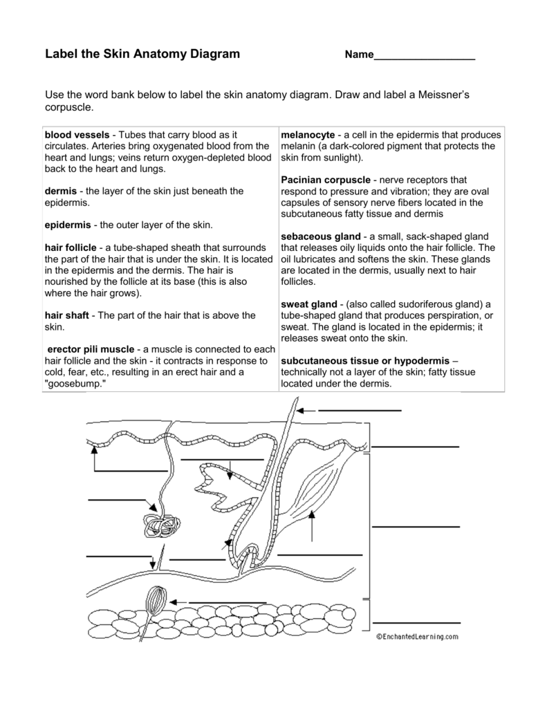
Label Skin Diagram Printout - EnchantedLearning.com Label Skin Anatomy Diagram Printout.

skin diagram coloring and labeling worksheet us hair ...
Skin: Layers, Structure and Function - Cleveland Clinic Skin. As the body's largest organ, skin protects against germs, regulates body temperature and enables touch (tactile) sensations. The skin's main layers include the epidermis, dermis and hypodermis and is prone to many problems, including skin cancer, acne, wrinkles and rashes. 216.444.5725. Appointments & Locations.

Vektor Stok Human Body Skin Anatomy Diagram Chart (Tanpa ...
Skin Diagram Labeling Skin Diagram Labeling. 1. Label the diagram with the letters below according to the structure/area they describe. You may label with a line or put the label ...
Skin Labeling Quiz
Labeled Skin Structure Diagram | Quizlet Epidermis Outermost layer of skin, provides a strong, waterproof, protective barrier for the body. Dermis Fibrous and elastic tissue, provides strength and elasticity to the skin and supports the epidermis, home to hair follicles, glands, nerves etc Papillary Layer

STRUCTURED/APPLICATION/SKILL TYPEGiven below is a ...
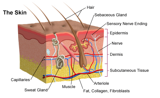
Anatomy of the Skin | SEER Training - National Cancer Institute The skin is the body's largest organ; covering the entire outside of the body, it is about 2 mm thick and weighs approximately six pounds. It shields the body against heat, light, injury, and infection. The skin also helps regulate body temperature, gathers sensory information from the environment, stores water, fat, and vitamin D, and plays a ...

Pin by Katlyn Mcdonough on Skin science | Skin anatomy ...
Label the skin - Teaching resources - Wordwall Label the Skin Game - Label the skin - Label the skin - Label the diagram of the skin - Label the skin (Level 3) - Skin diagram to label - The Skin.

Solved Integumentary System 10 points Label the diagram ...
Skin Labeling | Biology Game - Turtle Diary Identify and label figures in Turtle Diary's interactive online game, Skin Labeling! Drag the given words to the correct blanks to complete the labeling!
Label the skin - Teaching resources
Skin Diagram || How to draw and label the parts of skin - Pinterest Nov 10, 2022 - 'Skin Diagram || How to draw and label the parts of skin' is demonstrated in this video tutorial step by step.The sense of touch had received ...

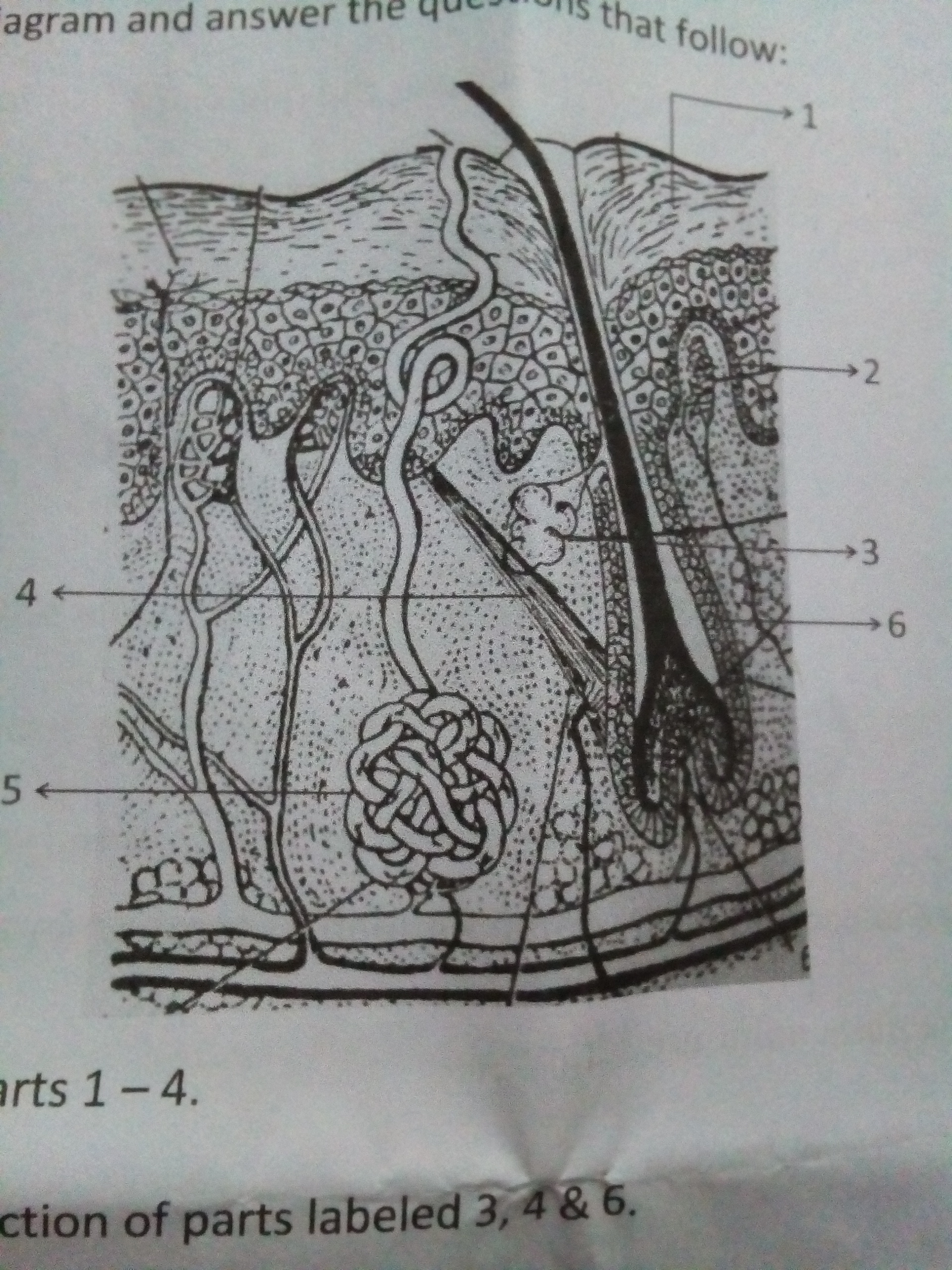
Activity Identify and name the parts labeled 1 through 7 in ...
Skin structure diagram - Teaching resources - Wordwall L3 skin diagram NEW Labelled diagram by Kmiller14 Skin structure Match up by Siobhan45 Skin structure Match up by Kellyrandall Level 3 Advanced Skin Diagram Labelled diagram by Lauragalgey Beauty Therapy Skin diagram to label Labelled diagram by Wyeoman Technical education Biology Skin Diagram (L3 camouflage) Labelled diagram by Kmiller14

label: skin diagram Diagram | Quizlet
Anatomy of the Epidermis with Pictures - Verywell Health The epidermis is composed of layers of skin cells called keratinocytes. Your skin has four layers of skin cells in the epidermis and an additional fifth layer in areas of thick skin. The four layers of cells, beginning at the bottom, are the stratum basale, stratum spinosum, stratum granulosum, and stratum corneum.

Anatomy 2017- Unit 2 Label Parts of Skin Diagram Diagram ...
Anatomy of the Skin - Stanford Children's Health The skin is made up of 3 layers. Each layer has certain functions: Epidermis. Dermis. Subcutaneous fat layer (hypodermis) ...
23,714 Skin Anatomy Stock Photos, Pictures & Royalty-Free ...
Integumentary System: Skin Diagram to Label

Human Body: The Skin
SKIN: Labeling diagram,Review / Research Worksheets| Digital Distance Learning
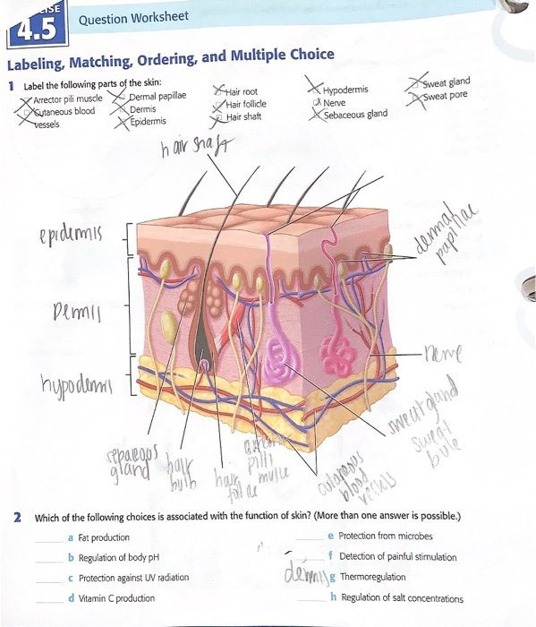
Solved 4.5 Label the following parts of the skin ***Which of ...

STRUCTURED/APPLICATION/SKILL TYPEGiven below is a ...

SKIN DIAGRAM Let's Label it Up!. SKIN Diagram - LEFT ...
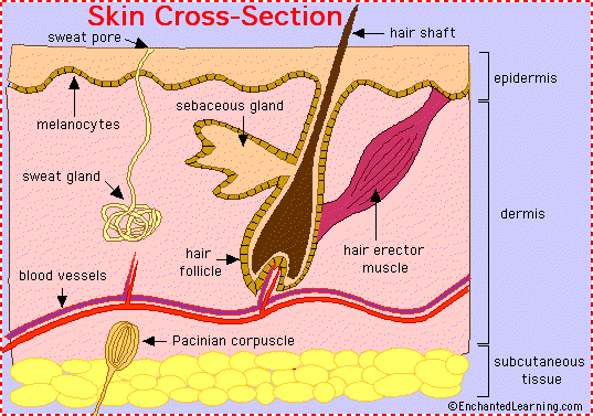
Skin Anatomy - EnchantedLearning.com

How to draw skin LS | Biology diagrams, Biology drawing ...
Skin Diagram Quiz questions & answers for quizzes and tests ...

Anatomy of human skin with labels Stock Photo - Alamy

Skin Labeling Quiz Diagram | Quizlet
Unit 4 Skin Labeling Quiz

File:Anatomy The Skin - NCI Visuals Online.jpg - Wikimedia ...
Human skin diagram hi-res stock photography and images - Alamy

Berkas:Diagram of human skin.jpg - Wikipedia bahasa Indonesia ...
Label the Skin Anatomy Diagram
Post a Comment for "40 skin diagram to label"