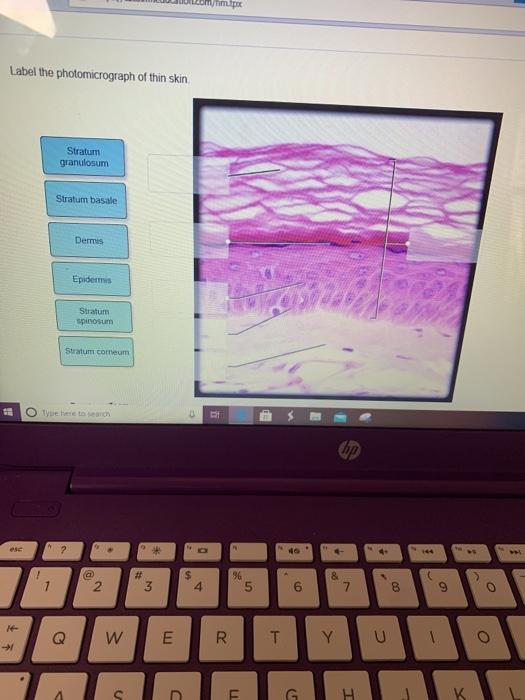
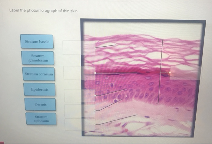
43 label the photomicrograph of thin skin
anatomy lab, exam 3, lab 9, Spinal Nerves, Integument, and ... - Quizlet Label the photomicrograph of thin skin. stratum corneum stratum granulosum stratum spinosum stratum basale dermis epidermis hypodermis the layer of skin beneath the dermis, which serves as a storage repository for fat Name the yellow highlighted structures that pass through the intervertebral foramina. spinal nerves Label The Photomicrograph Of Thin Skin And Its Accessory Structures ... Part a is a micrograph showing a cross section of thin skin. As a person ages, the melanin production decreases, and hair tends to lose its color and becomes gray and/or white. Label the photomicrograph of the skin and its accessory structures. The skin and its accessory structures make up the integumentary system,.
Label The Photomicrograph Of Thin Skin. - Skin Model 1 - YouTube Label the photomicrograph of thin skin. It has a fifth layer, called the stratum lucidum, located between the stratum corneum and the stratum granulosum ( (figure)). Hair sebaceous gland dermis hair follicle epidermis duct of sebaceous. Label the photomicrograph of thin skin. Thin skin versus thick skin.
Label the photomicrograph of thin skin
Figure 7.1: Photomicrograph of Skin Diagram | Quizlet Start studying Figure 7.1: Photomicrograph of Skin. Learn vocabulary, terms, and more with flashcards, games, and other study tools. Bio Lab Chapter 6 Quiz Flashcards | Quizlet Identify the type of tissue that composes the epidermis of the skin. stratified squamous epithelial tissue Identify the structures of the dermis. dense connective tissue with fibers oriented in many directions dense irregular loose connective tissue characterized by long, thin dark fiber areolar tissue › homework-help › questions-andSolved Label the photomicrograph of thick skin | Chegg.com Label the photomicrograph of thick skin This problem has been solved! You'll get a detailed solution from a subject matter expert that helps you learn core concepts. See Answer Question: Label the photomicrograph of thick skin Show transcribed image text Expert Answer 92% (12 ratings) Transcribed image text: Label the photomicrograph of thick skin
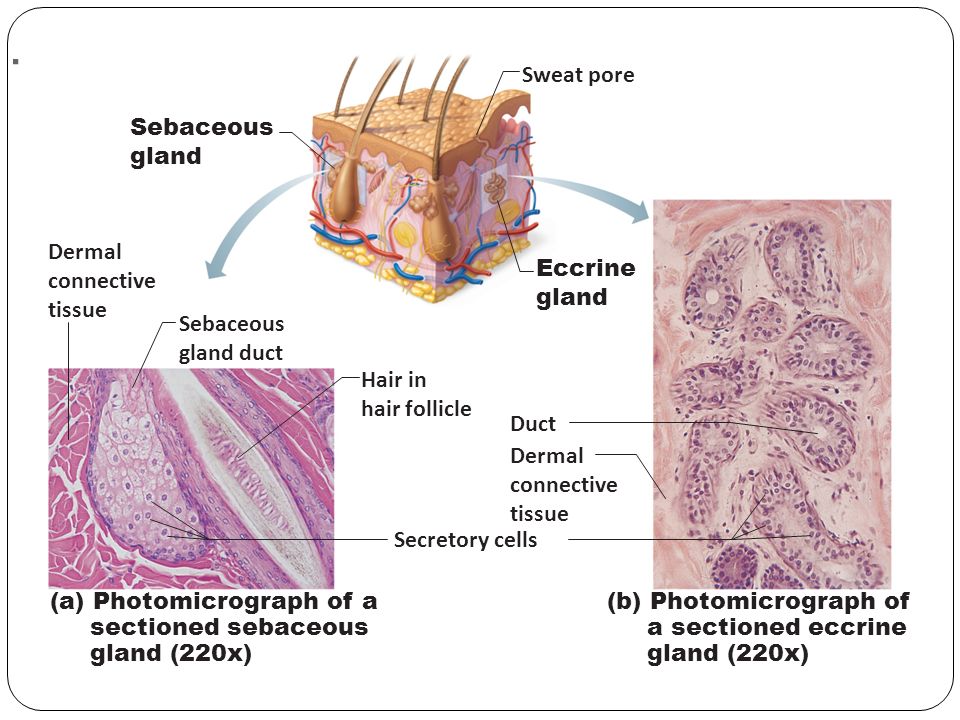
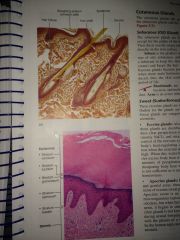
Label the photomicrograph of thin skin. photomicrograph of thick skin Diagram | Quizlet photomicrograph of thick skin Diagram | Quizlet photomicrograph of thick skin + − Learn Test Match Created by mckennawebber Terms in this set (7) epidermis (stratum corneum - stratum basale) ... stratum corneum ... stratum lucidum ... stratum granulosum ... stratum spinosum ... stratum basale ... dermis ... Final Exam A&P 1 Flashcards | Quizlet Label the structures of the skin and subcutaneous tissue. Label the photomicrograph of thick skin. Epidermis, stratum corneum, stratum lucidum, stratum granulosum, stratum spinosum, stratum basale, dermis Label the photomicrograph of thin skin Hair shaft, epidermis, dermal root sheath, sebaceous gland, dermis, hair matrix Label The Photomicrograph Of Thick Skin - Lichen 4 The Algal Layer ... Label the photomicrograph of thick skin. Thick skin · stratum basale (also known as s. Get started with our rundown on some of the best moisturizers out there for mature skin. "thick skin" is found only on the palms of the hands and the soles of the feet. Thin skin versus thick skin. Start studying photomicrograph of thick skin. Unit 4 lab.docx - LAB Unit 4 EXERCISE 7: The Integumentary... FIGURE 7.2: Photomicrograph of the skin. epidermis (EPI-derm-is) • dermal papillae (puh-PILL-ee) • hypodermis (HY-poh- der-mis) • papillary (PAP-il-lary) layer of dermis • reticular layer of dermis 1. Dermal Papillae 2. Epidermis 3. Papillary layer of dermis 4. Reticular layer of dermis 5. Hypodermis
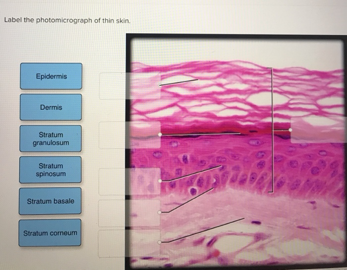
› game › photomicrograph-ofPhotomicrograph of Thin Skin Quiz - PurposeGames.com This is an online quiz called Photomicrograph of Thin Skin. There is a printable worksheet available for download here so you can take the quiz with pen and paper. Quiz Points. 5 p. You need to get 100% to score the 5 points available. Game of the Day. Stars of Astronomy: the 20 Greatest Astronomers. by Wentu. 2,330 plays. Label The Photomicrograph Of Thick Skin. - Martina Eisenhower 1 answer to label the photomicrograph of thin skin. The epidermis, made of closely packed epithelial cells, and the dermis, made of dense, irregular connective tissue . Epidermis Of Thick Skin from eugraph.com The skin is composed of two main layers: Thick skin showing epithelial detail. Practice labeling the layers of the skin. Label the photomicrograph of thin skin. O Stratum granulosum O Stratum ... Label the photomicrograph of thin skin. Stratum granulosum Stratum spinosum Dermis Epidermis Stratum corneum Stratum basale We store cookies data for a seamless user experience. brainly.com › question › 30088019Label the photomicrograph of thin skin. Dermis Duct of ... Dec 27, 2022 · The skin is the largest organ in the body; it extends from the inside of the body to the outside, has a thickness of about two millimeters, and weighs around six pounds on average. It protects the body against excessive heat and light, as well as injury and infection.
Label The Photomicrograph Of Thick Skin Quizlet : Organs And Structures ... Label the photomicrograph of thin skin. Start studying photomicrographs of skin (thick skin). Common causes range from illness to injury to inflammation. Chapter 6 Study Set Flashcards Practice Test Quizlet from quizlet.com Learn vocabulary, terms, and more with flashcards, games, and other study tools. Skin discoloration, defined by healthline ... › homework-help › questions-andSolved Label the photomicrograph of thin skin. Dermis Duct ... Label the photomicrograph of thin skin. Dermis Duct of sebaceous gland Hair Follicle Sebaceous gland Hair Epidermis This problem has been solved! You'll get a detailed solution from a subject matter expert that helps you learn core concepts. See Answer Question: Label the photomicrograph of thin skin. Photomicrograph of Thick Skin Quiz - PurposeGames.com Label a Nephron. Science. English. Creator. ninalahoti. Quiz Type. Image Quiz. Value. 16 points. Likes. 47. Played. 103,027 times. Printable Worksheet. Play Now. ... This is an online quiz called Photomicrograph of Thick Skin. There is a printable worksheet available for download here so you can take the quiz with pen and paper. open.oregonstate.education › aandp › chapter5.1 Layers of the Skin – Anatomy & Physiology Skin that has four layers of cells is referred to as "thin skin.". From deep to superficial, these layers are the stratum basale, stratum spinosum, stratum granulosum, and stratum corneum. Most of the skin can be classified as thin skin. "Thick skin" is found only on the palms of the hands and the soles of the feet.
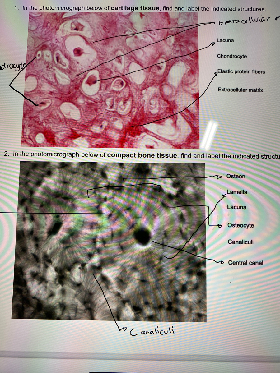
Answered: 1. In the photomicrograph below of… | bartleby
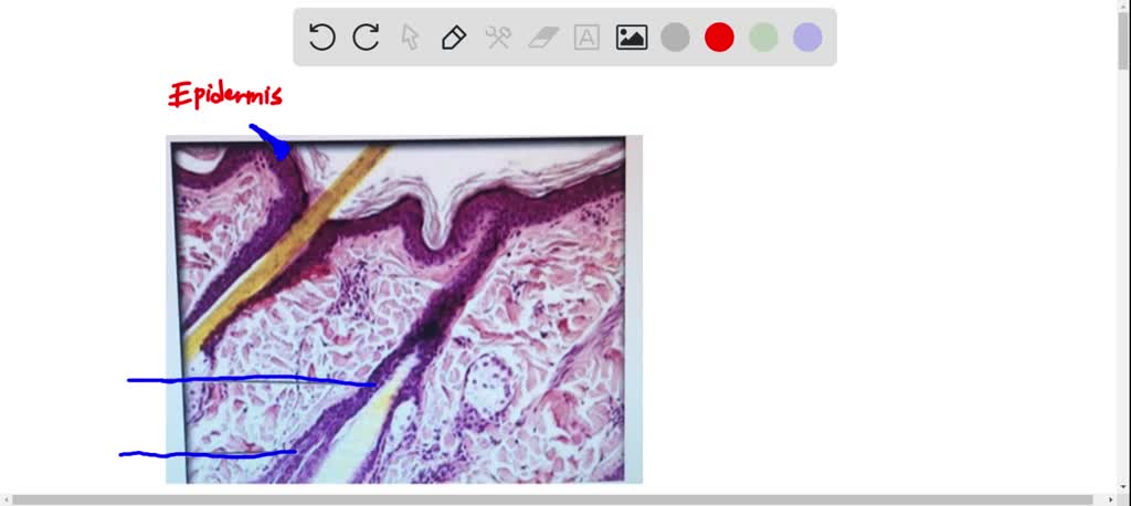
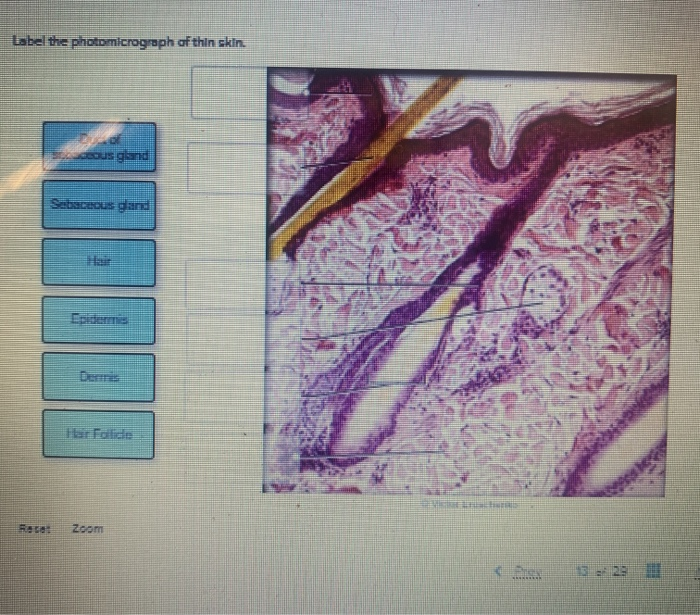
Sebaceous Gland Label The Photomicrograph Of Thin Skin - Blogger Label the photomicrograph of thin skin. Part a is a micrograph showing a cross section of thin skin. Name the 4 layers of thin skin in both the cartoon and the photomicrograph. This problem has been solved! Dermis duct of sebaceous gland hair follicle sebaceous gland hair epidermis.

BIOL 319 Lab 1 Flashcards | Quizlet
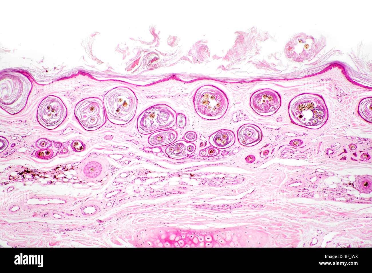
Skin overview 3 | Digital Histology Thick skin. Skin can be classified as either thick or thin, depending on the thickness of the epidermal layer. This image compares a diagrammatic representation of thick skin with a photomicrograph of a hematoxylin and eosin-stained section of primate skin. 200x.

View Image
Label the photomicrograph of thin skin. Dermis Duct of...ask 3 Answer of Label the photomicrograph of thin skin. Dermis Duct of sebaceous gland Hair Follicle Sebaceous gland Hair Epidermis

BIOL 319 Lab 1 Flashcards | Quizlet
Solved Label the photomicrograph of thin skin | Chegg.com Label the photomicrograph of thin skin This problem has been solved! You'll get a detailed solution from a subject matter expert that helps you learn core concepts. See Answer Question: Label the photomicrograph of thin skin Show transcribed image text Expert Answer 100% (25 ratings) Transcribed image text: Label the photomicrograph of thin skin

Ch. 22 Assessment Flashcards | Quizlet
photomicrographs of thin skin Flashcards | Quizlet photomicrographs of thin skin. Term. 1 / 4. stratum corneum. Click the card to flip 👆.
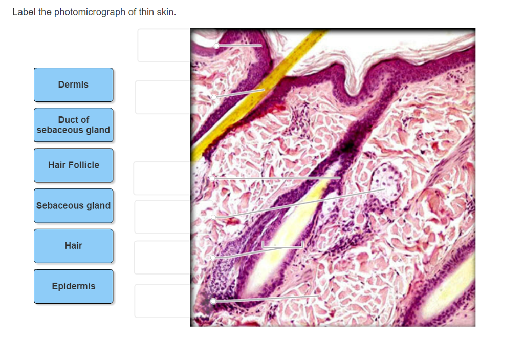
Solved Label the photomicrograph of thin skin. Dermis Duct ...
Lab 9: Pre-Lab Homework Flashcards | Quizlet Label the photomicrograph of thin skin. In general, nerves from the posterior division of the brachial plexus tend to innervate muscles that extend the parts of the upper limb. True/False True Match the label to its appropriate spinal cord component. -dorsal root ganglion -white matter -gray matter -epidural space -dorsal ramus
Solved Label the photomicrograph of thin skin. Epidermis ...
› questions › label-the(Solved) - Label the photomicrograph of thin skin. Label the ... Aug 5, 2021 · Label the photomicrograph of thin skin 1 Approved Answer Mohinee k answered on August 05, 2021 4 Ratings ( 9 Votes) Label the photomicrograph of the skin A photograph taken with the help of microscope . Skin is the largest sensory organ in body.its protect the body sense pain... solution .pdf

A&P Unit 2 Skin Tissue (Model, Photomicrographs & Graphic ...
Question : Question 31 points Label the photomicrograph of thin skin ... Question 31. A first grade teacher wishes to "shape" her student's writing of the alphabet. The teacher should: a, reward the child whenever the c... Question 31. A neurotransmitter that allows sodium ions to leak into a postsynaptic neuron causes: A) inhibitory postsynaptic damage to the myelin sheath C) excitatory postsynaptic...

Page 3 | 400+ Photomicrograph Pictures
courses.lumenlearning.com › layers-of-the-skinLayers of the Skin | Anatomy and Physiology I - Lumen Learning Most of the skin can be classified as thin skin. “Thick skin” is found only on the palms of the hands and the soles of the feet. It has a fifth layer, called the stratum lucidum, located between the stratum corneum and the stratum granulosum (Figure 2). Figure 2. Thin Skin versus Thick Skin.
Label tne photomicrograph Of the Skin and Its accessory structures, Sebaceous gland, Duct ofl, sebaceous gland, Epidermis, Hair follicle
› homework-help › questions-andSolved Label the photomicrograph of thick skin | Chegg.com Label the photomicrograph of thick skin This problem has been solved! You'll get a detailed solution from a subject matter expert that helps you learn core concepts. See Answer Question: Label the photomicrograph of thick skin Show transcribed image text Expert Answer 92% (12 ratings) Transcribed image text: Label the photomicrograph of thick skin
Simple ciliated columnar hi-res stock photography and images ...
Bio Lab Chapter 6 Quiz Flashcards | Quizlet Identify the type of tissue that composes the epidermis of the skin. stratified squamous epithelial tissue Identify the structures of the dermis. dense connective tissue with fibers oriented in many directions dense irregular loose connective tissue characterized by long, thin dark fiber areolar tissue
Solved met Label the photomicrograph of thin skin Stratum ...
Figure 7.1: Photomicrograph of Skin Diagram | Quizlet Start studying Figure 7.1: Photomicrograph of Skin. Learn vocabulary, terms, and more with flashcards, games, and other study tools.

Hair shaft Dermal papillae Epidermis Subpapillary vascular ...
Skin and the Integumentary System

a): A photomicrograph of the section of thin skin tissue from ...
Hair shaft Dermal papillae Epidermis Subpapillary vascular ...
Animals | Free Full-Text | Naturally Produced Lovastatin ...

Pin by nico x. on Anatomy | Games, Tetris, Anatomy
Integumentary System Overview
Connective Tissue Lab
Skin cross section hi-res stock photography and images - Alamy

Solved Label the photomicrograph of thin skin | Chegg.com

Fingernail Anatomy Picture Image on MedicineNet.com
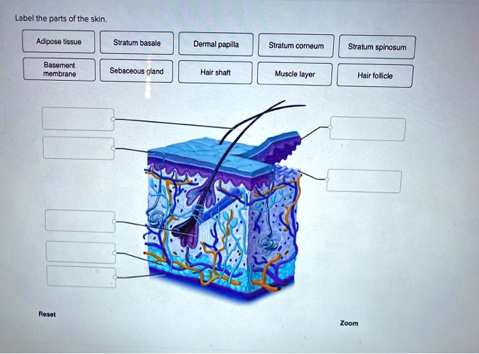
SOLVED: Label tne parts of the skin: Adipose tissue Stratum ...

Histologia de la piel parte II

Stratum Lucidum 5 Translucent layer found in thick skin ...

The Microscopic Beauty of Plants and Trees by Robert Berdan ...

a) A photomicrograph of the section of thin skin tissue from ...
Solved Label these structures located in axillary skin. Hair ...
Epidermis of onion (Allium cepa) with cells, nucleus and ...

Supraclavicular extra-renal angiomyolipoma: a challenging ...

Wound Healing Properties and Antimicrobial Effects of Parkia ...
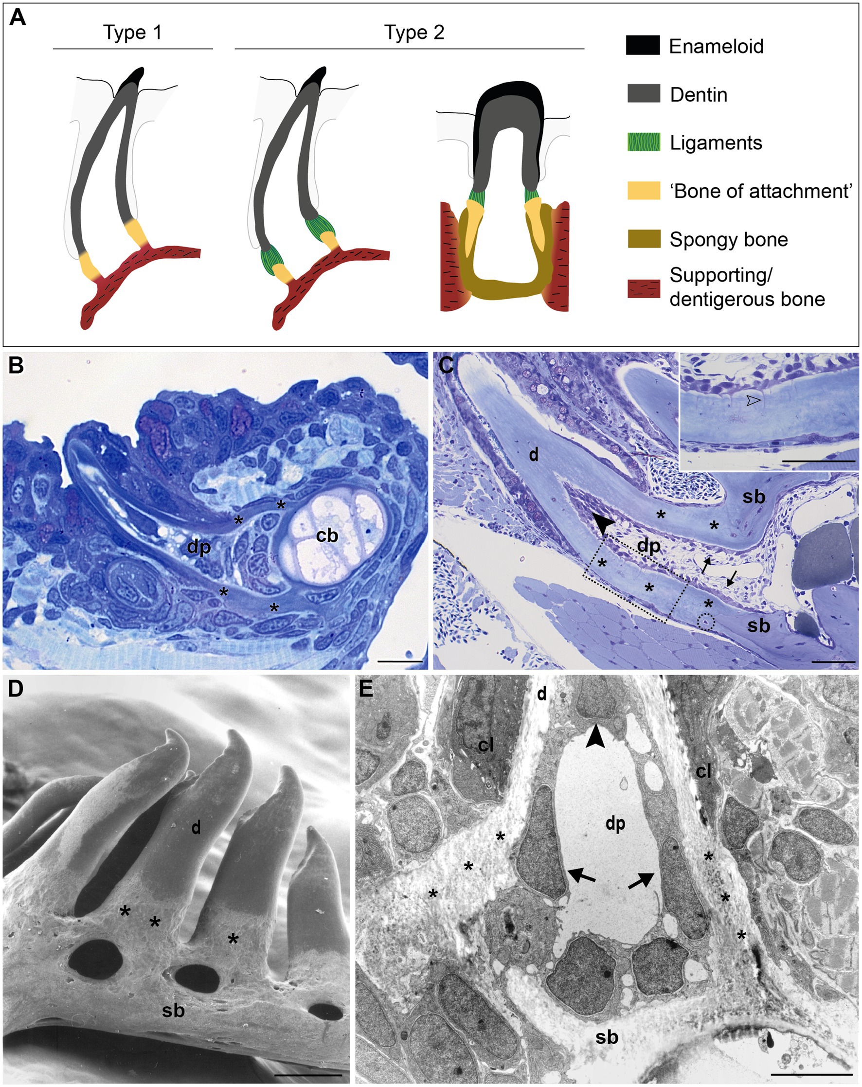
Frontiers | Cells at the Edge: The Dentin–Bone Interface in ...
Solved Label the photomicrograph of thin skin. deous gland ...

Primary Liver Cancers—Part 1: Histopathology, Differential ...

unit 4 lab.docx - LAB Unit 4 EXERCISE 7: The Integumentary ...

SciELO - Brasil - Giant epidermal inclusion cyst of the ...

Endocrine System Anatomy And Physiology - Human Anatomy ...

Hair | Biology for Majors II

BIOLOGY - microscopia.info
Integumentary System Overview
Anatomy Lab Practical 1 Flashcards - Cram.com

Ch 27 Lab Flashcards | Quizlet
Post a Comment for "43 label the photomicrograph of thin skin"